EclipseXRM-900 - High Resolution X-ray Microscope
Computed TomographyComputed Tomography - Life SciX-ray Microscopy
Submicron resolution even at large working distances

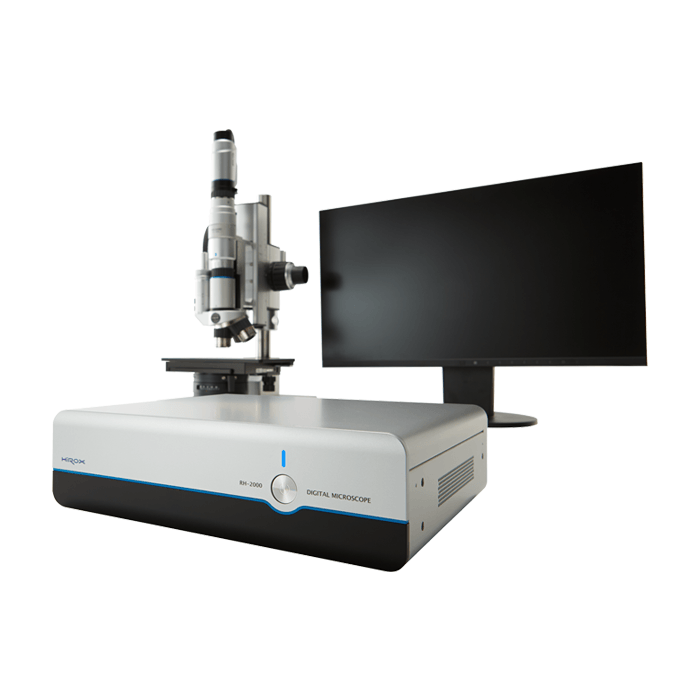
Highly Customizable digital microscope for 2D and 3D examination and analysis of samples from 0x to 10,000x magnification