
This whitepaper discusses the use cathodoluminescence of sedimentary rocks, specifically sandstone.

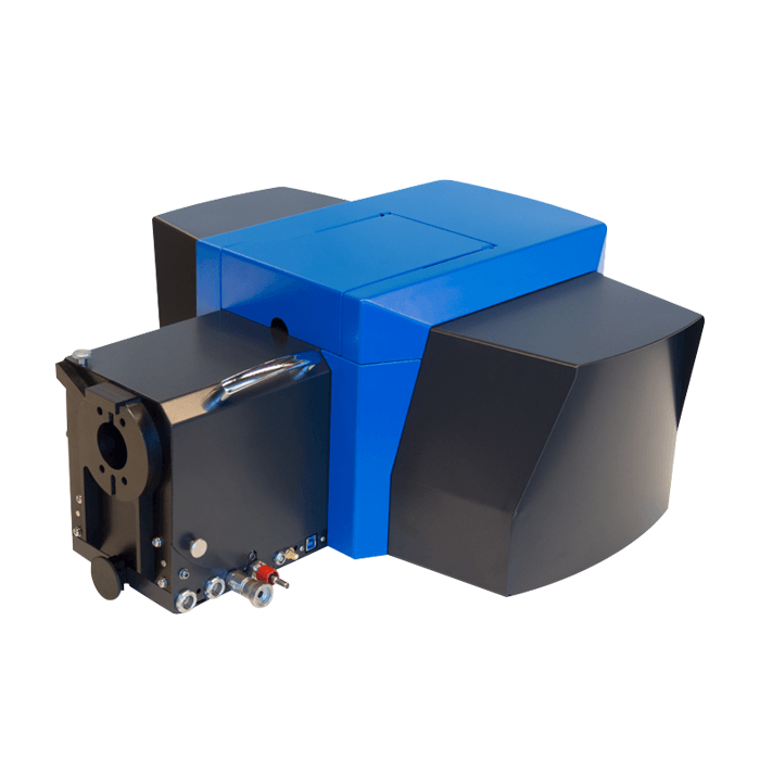
The SPARC is a high-performance cathodoluminescence detector, which is unique for its modular design, ease-of-use, multiple imaging modes and the ability to be retrofitted on any scanning electron microscope (SEM).
The various cathodoluminescence analytical modes afforded by the DELMIC SPARC are summarised below. For more details on how they work, read our article on Recent Advances in Cathodoluminescence Imaging Modes or visit Cathodoluminescence Imaging Modes and Applications for a collection of videos and webinars that will provide yet more details.
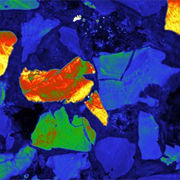
A fast analog PMT detector can be used for large-scale imaging. This allows for the rapid inspection of large areas, ideal for geological applications, fast device inspection, and efficient region-of-interest finding. A filter wheel can be used for spectral differentiation.
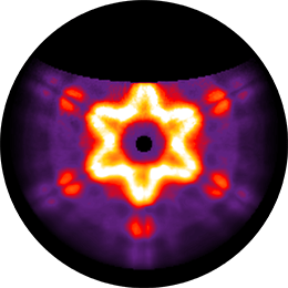
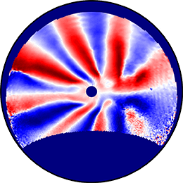
The SPARC provides the unique option to acquire angle-resolved images. Rather than focusing the light signal on a fiber or narrow opening, an image of the mirror can be projected onto an imaging camera. This allows for the detection of the directionality of the emitted light, also known as momentum spectroscopy. In this mode, a filter wheel is used to spectrally distinguish the different emission wavelengths.
When the SPARC system is used in spectral mode, the light coming from the mirror is focused on a slit or fiber connected to a Czerny-Turner spectrograph. A variety of imaging detectors can be used to cover a spectral range of 200-1600 nm. By scanning the e-beam across the sample, a spatially resolved hyperspectral image is produced.
Using a polariser or even a full polarimeter in the angle-resolved mode allows for the reconstruction of the polarisation state (Stokes vector) of CL for different emission angles. An advanced correction for the optical system including the paraboloid mirror is required for this reconstruction. This is provided with the polarisation system.
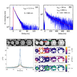
With a Lab Cube module, an add-on for the standard SPARC spectral system, it is now possible to perform g(2) and lifetime imaging and observe the time dynamics of various nanomaterials. Time-resolved imaging is highly relevant for a wide range of applications, including semiconductors for photovoltaics and light-emitting devices, as well as single emitters for quantum information processing and sensing.
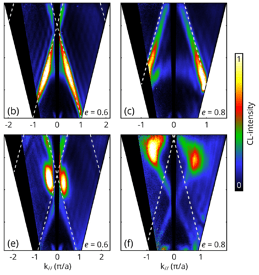
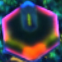
Energy-Momentum Cathodoluminescence Imaging is a new technique which can be applied to track the directionality through energy and momentum space with very high precision. It is a great tool for mapping the optical properties of a wide range of dispersive and anisotropic systems, paving the way for a broad range of studies on complex nanophotonic systems.


