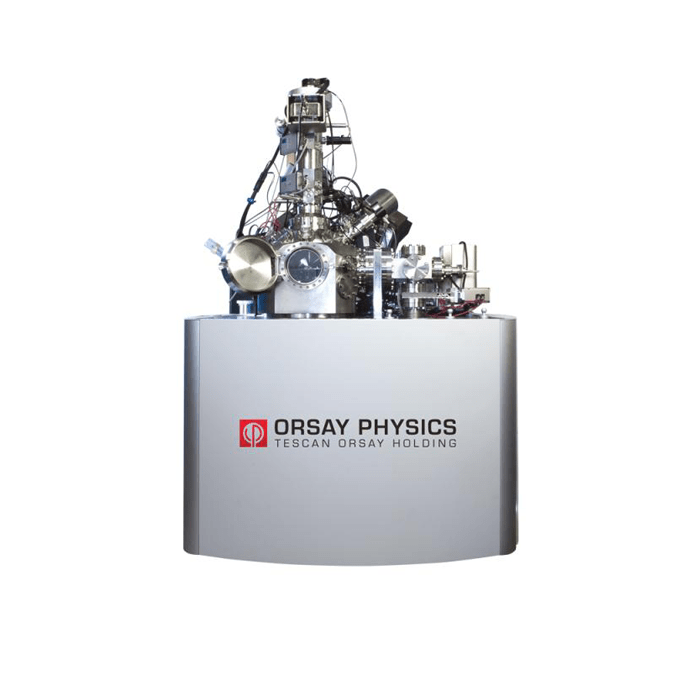
METEOR

Integrated top down cryo-CLEM imaging system

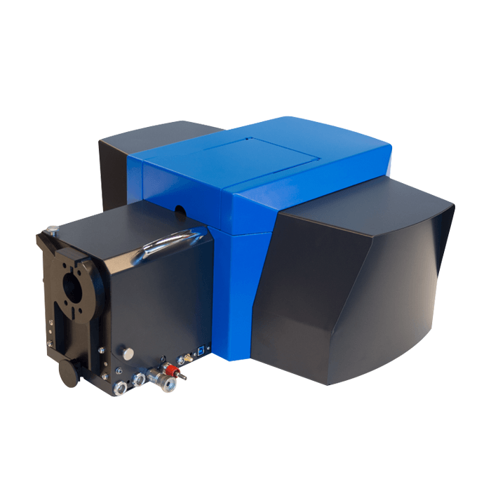
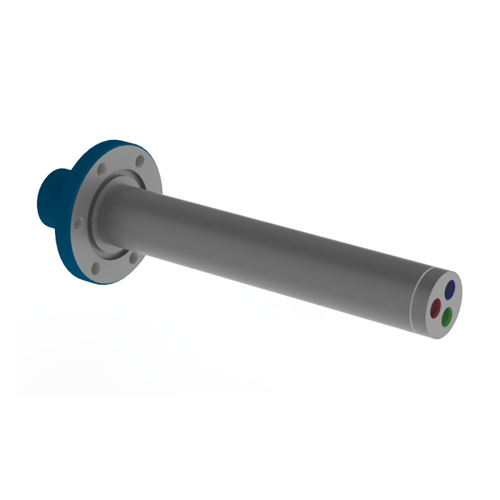
METEOR is the world’s first and only fully integrated solution to optimise the cryo-ET workflow by reducing complicated transfer steps and increasing sample yield
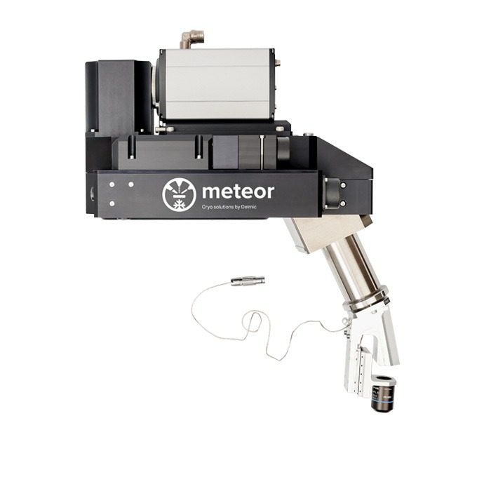
  • delmic meteor

    Key Features

    • FM enables region of interest (ROI) finding within the FIB/SEM chamber and therefore better lamella targeting
    • Integrated cryo CLEM allows for better XY correlation as the same sample stage and coordinates are used
    • Provides the ability to inspect lamellae for ROI inclusion during and after milling. Take only the useful lamellae to the cryo TEM.
    • Removing the transfer steps between LM and SEM reduces the risks of sample damage (devitrification/mechanical) and ice contamination
    • FM imaging in a high vacuum environment means lower ice deposition rate compared with ambient conditions
    • Easily adaptable to the users’ current workflow (plunge freezing/ HPF)

Integrated Top-Down Cryo-CLEM Imaging System

METEOR is the world’s first and only fully integrated solution to optimise the cryo-ET workflow by reducing complicated transfer steps and increasing sample yield.

METEOR 2.0 – Released March 2025

METEOR 2.0, the next evolution of Delmic’s integrated cryo-fluorescence microscope (cryo-FM) designed to simplify and accelerate your cryo-electron tomography (cryo-ET) workflow. It enhances researchers ability to confidently identify and target regions of interest—without added contamination risk or workflow delays.

New Features in METEOR 2.0

  • 8x larger field of view – making it easier to navigate and identify ROIs
  • Advanced filter wheel – new high-speed, high-capacity filter wheel supports up to six customisable single band-pass emission filters, along with reflection imaging. It minimises spectral bleed-through for cleaner multi-channel imaging and more accurate signal interpretation. Faster switching between channels also boosts imaging throughput, increasing workflow efficiency
  • Optimised optical path – optimised photon detection while minimising signal loss. In combination with high NA-objectives and high QE cameras, METEOR 2.0 allows you to measure the weakest fluorescence signals using the minimal excitation power. This also minimised devitrification risk and maximises confidence your region of interest is captured in the final lamella.
  • Software integration – fully integrated ODEMIS software ensures seamless operation by integrating imaging, correlation, and automation functionalities. It supports easy data import and compatibility with external software, enhancing workflow efficiency.

METEOR 2.0’s new features further enhance your ability to successfully perform cryo-ET imaging and to “see more with less” by providing a system that reduces contamination, saves time, and enhances imaging capabilities.

  • Delmic Meteor 7 step simplified workflow

    Optimised Cryo-ET Workflow – Just 7 Steps

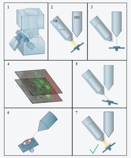
    METEOR can reduce the complexity of cryo-correlated light and electron microscopy (CLEM) workflow greatly by removing many transfer steps in the current laborious lamella preparation workflow. Traditional workflows can include as many as 22 steps. METEOR reduces this to just 7 fabrication steps:
    1. Load your sample
    2. Capture FLM image
    3. SEM image without sample transfer
    4. Directly move to the region of interest (ROI) after correlating the two images
    5. Mill your lamella
    6. Confirm the presence of ROI with FM
  • Delmic meteor cryo-ET applications

    Applications

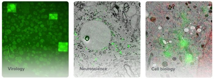
    METEOR has a range of application in life science/biological imaging including:
    • Virology
    • Neuroscience
    • Cell Biology
  • Introduction to the METEOR

    METEOR, the first commercially available integrated fluorescence microscope module, METEOR will smash the benchmark of the highest productivity in cryo electron tomography (cryo-ET) workflow.

  • All
  • AFM/SPM/SNOM
  • Automated Mineralogy
  • CL
  • CLEM
  • EBSD
  • EDS
  • Electron Beam Lithography (EBL)
  • Electron Microscopy
  • Fabrication
  • FIB
  • Hyperspectral
  • In situ
  • Micro XRF
  • Microscopy
  • Raman
  • SEM
  • Spectroscopy
  • TEM
  • Thermal Probe Lithography
  • WDS