
EclipseXRM-900 - High Resolution X-ray Microscope
Computed TomographyComputed Tomography - Life SciX-ray Microscopy
Submicron resolution even at large working distances


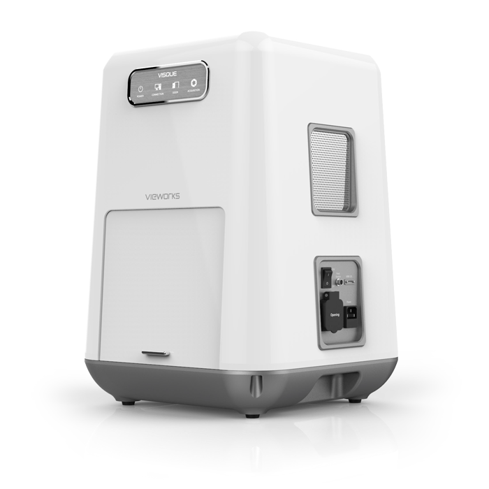
The Vieworks VISQUE® InVivo Smart LF is a compact in vivo optical imaging system that offers fluorescent and luminescent detection from green to near-infrared and also time-lapse acquisition as fast as 37 frames per second.
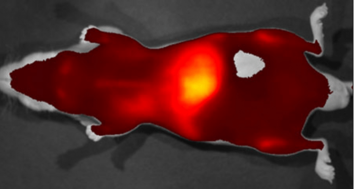
Nude mouse image taken 2 days after Exosome-NIR dye complex injection through the tail vein.


