TESCAN CLARA

The only UHR-SEM in the market that doesn’t discriminate samples

Field-free analytical UHR SEM for materials characterisation at nanoscale

  • Key Features

    • Largest field of view – Up to 15mm @ WD 10mm powered by TESCAN Wide Field Optics™
    • Fast imaging condition switching – Seamless navigation from mm to nm scales
    • Simultaneous UHR SE & BSE imaging - Up to 6 signals, producing enhanced contrast for deeper insights
    • True field-free UHR imaging at low keV - 1nm @1kV ideal for charging, sensitive and magnetic samples
    • Integrated analytical workflows – Open platform supporting EDS, EBSD, Raman, and automation
    • MultiVac low-vacuum imaging – Automated operation with water vapor and large FOV

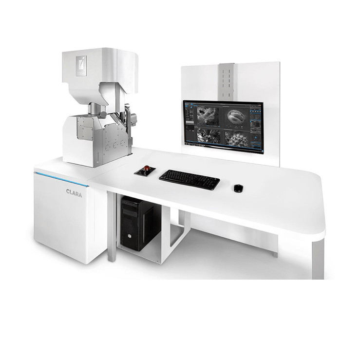
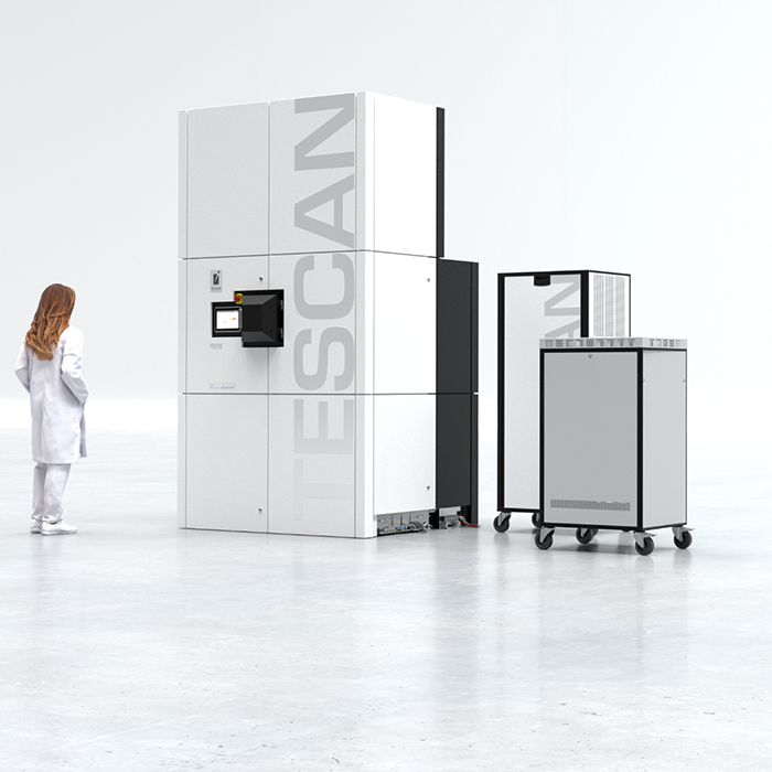
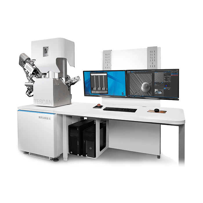
The new TESCAN CLARA™ FEG-SEM is an ultra-high-resolution field-emission scanning electron microscope designed for advanced materials characterisation. Combining TESCAN’s BrightBeam™ field-free electron optics, advanced SE and BSE detection, low-energy imaging and automated workflows, CLARA is designed to extract more information from challenging samples while reducing the compromises traditionally associated with high-resolution SEM imaging.

With resolution down to 0.7 nm at 30 keV and 1.1 nm at 500 eV, CLARA delivers high-resolution imaging across a broad range of accelerating voltages. Beam deceleration can further achieve 1.0 nm at 1 keV, providing researchers with high-resolution surface imaging at very low landing energies.

Key Capabilities

Ultra-high-resolution imaging at low landing energies

One of CLARA’s major strengths is its ability to maintain high-resolution imaging at low accelerating voltages.
The system delivers:

High vaccum mode

  • 1.0 nm @ 1keV, BDT
  • 1.1nm @ 0.5keV
  • 0.7 nm @ 15 keV
  • 0.7 nm @ 30 keV, STEM

Low vacuum mode

  • 2.0 nm @ 30 keV (BSE)
  • 1.1 nm @ 30 keV with GSD
  • 2.5 nm @ 3keV with GSD

This low-kV capability is particularly valuable when examining surface structures, nanoparticles, thin films, beam-sensitive materials and non-conductive samples, where higher electron energies can increase interaction volume, charging or beam damage.

Why this matters

High-resolution imaging isn’t simply about achieving the smallest possible resolution specification. For many materials scientists, the ability to maintain useful resolution at low landing energies is more important because it allows the microscope to interrogate the surface and near-surface region with greater sensitivity.

BrightBeam™ Field-Free Electron Optics

At the heart of CLARA is TESCAN’s BrightBeam™ field-free UHR electron column.
The field-free architecture is particularly useful when working with magnetic materials, because the sample is not subjected to the strong magnetic field associated with conventional immersion-lens configurations.

This allows researchers to examine magnetic specimens while maintaining high-resolution imaging capability.

The combination of BrightBeam optics and beam deceleration also allows CLARA to operate effectively at very low landing energies, supporting imaging of delicate and challenging materials.

Multi-Contrast Detection: More Information from Every Scan

Resolution is only one part of an SEM’s performance.
The new CLARA is designed around the principle that different electron signals reveal different aspects of a material.
Its detection system combines:

  • Axial detector
  • MultiDetector™
  • Energy-filtered BSE detection
  • Angular/angle-selective BSE contrast
  • Secondary electron imaging
  • Additional BSE detection options

This allows users to obtain complementary information about:

  • Surface morphology
  • Topography
  • Material composition
  • Phase differences
  • Near-surface structures
  • Thin layers
  • Contamination
  • Buried features

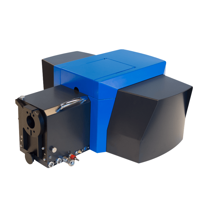
TESCAN’s MultiDetector can perform energy-filtered BSE imaging, allowing researchers to select different portions of the BSE signal to emphasise particular material contrasts.
The Multi-Contrast Detection Advantage

Rather than repeatedly imaging the same region using different detector configurations, CLARA is designed to acquire complementary SE and BSE information from the same region of interest.
That can reduce the need for rescanning, repositioning and repeated measurements—particularly valuable when working with beam-sensitive samples or when precise correlation between signals is required.

Characterise Difficult Samples Using MultiVac

CLARA is particularly well suited to samples that can be problematic for conventional high-resolution SEMs.
These include:

  • Non-conductive materials
  • Magnetic materials
  • Beam-sensitive specimens
  • Porous materials
  • Outgassing materials
  • Highly topographic surfaces
  • Nanomaterials
  • Thin films
  • Polymers
  • Energy materials

The MultiVac™ system provides low-vacuum operation using either nitrogen or water vapour, with operation up to 500 Pa. This expands the range of specimens that can be examined without relying on conventional conductive coatings or extensive sample preparation.

The ability to use water vapour in low-vacuum imaging is particularly interesting because it can improve signal quality and secondary electron contrast compared with conventional low-vacuum conditions.

Wide Field Optics®: From Overview to Nanometres

One of the frustrations of high-resolution SEM operation can be navigating from a large sample area to a nanoscale region of interest.

TESCAN’s Wide Field Optics® is designed to provide seamless navigation from 1× magnification through to 2,000,000×, allowing users to maintain spatial context while moving between overview and high-resolution imaging.
This is particularly useful for:

  • Locating defects
  • Navigating large samples
  • Finding specific particles or structures
  • Correlating low- and high-magnification observations
  • Moving rapidly between multiple regions of interest

Automation and Reproducibility

CLARA isn’t designed solely for experienced SEM specialists.

TESCAN has incorporated a range of automated tools intended to reduce setup time and improve reproducibility between operators as well as minimising time to data.
These include:

  • In-Flight Beam Tracing® – Automatically optimises electron-beam conditions to help users reach appropriate imaging conditions quickly
  • Automated focus and stigmation – Reduces the amount of manual adjustment required to achieve high-quality images
  • Essence™ software – Provides the operating environment for imaging, instrument control and workflow management
  • Essence™ 3D Collision Model – Provides real-time visualisation of the chamber and sample/stage configuration to help prevent collisions
  • VisualCoder™ – Provides no-code automation for custom imaging workflows
  • FIB-SEM Expert PI – Provides Python-based scripting capabilities for advanced and automated workflows

For core facilities and multi-user laboratories, these capabilities are particularly valuable because they help reduce operator-to-operator variability, abbreviate the operator training process and streamline workflows.

Large Chamber and Analytical Flexibility

CLARA is not simply a high-resolution imaging instrument.
Its large chamber is designed to accommodate a broad range of analytical and in-situ techniques, with 20 chamber ports available for detector and accessory integration. The system can accommodate samples up to approximately 335 × 280 × 133 mm and up to 8 kg, depending on configuration.

Available or integrable analytical techniques include:

  • EDS
  • EBSD
  • Raman spectroscopy
  • Cathodoluminescence
  • WDS
  • ToF-SIMS
  • 4D-STEM
  • In-situ mechanical testing
  • Heating
  • Tensile testing

This makes CLARA particularly attractive as a research platform rather than simply an imaging SEM.

Key Applications

Suited to multi-user and central facilities, CLARA is ideally suited to applications such as:

  • Advanced Materials – Investigate nanoscale surface morphology, phases, interfaces, defects and microstructure in advanced materials
  • Battery Materials – Low-kV imaging and advanced BSE contrast can be used to investigate electrode materials, separators, particle degradation, interfaces and SEI-related structures. TESCAN specifically positions CLARA for battery R&D, quality control and failure analysis
  • Semiconductor Research – The combination of high-resolution imaging, low landing energies and analytical integration makes CLARA suitable for nanoscale device and materials characterisation
  • Metals and Alloys – Use high-resolution SE/BSE imaging alongside EDS and EBSD to investigate grains, phases, inclusions, interfaces and surface defects
  • Nanomaterials – CLARA’s low-kV performance is particularly relevant to nanoparticles, nanostructured surfaces and other materials where surface-sensitive information is critical
  • Polymers and Beam-Sensitive Materials – Low-energy imaging and MultiVac operation can help minimise charging and beam damage while retaining useful surface detail
  • Surface Modifications and Coatings – Low landing energies and energy-filtered BSE imaging provide tools for investigating thin layers, coatings, contamination and near-surface material variations.

Reasons to Consider CLARA

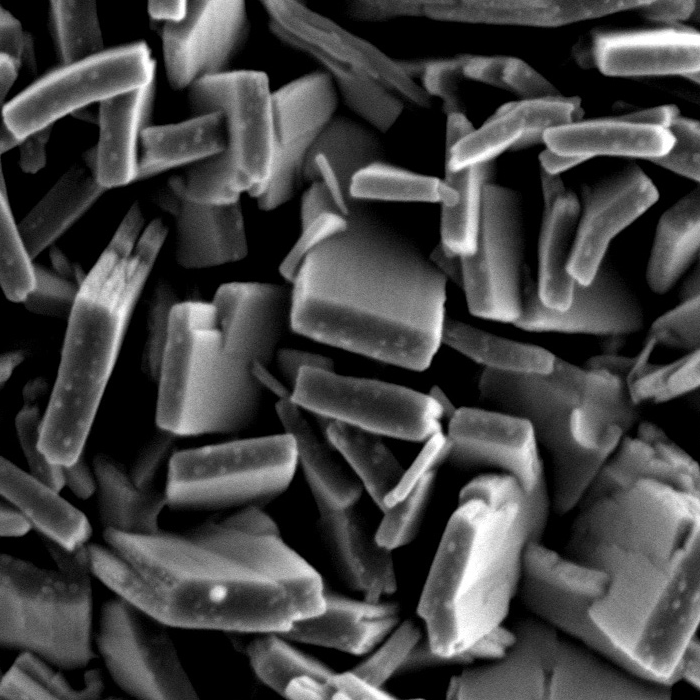
UHR at low keV resolves nanoparticle featuress without beam damage or charge artifacts on the carrier.
Mesoporous silica SBA-15

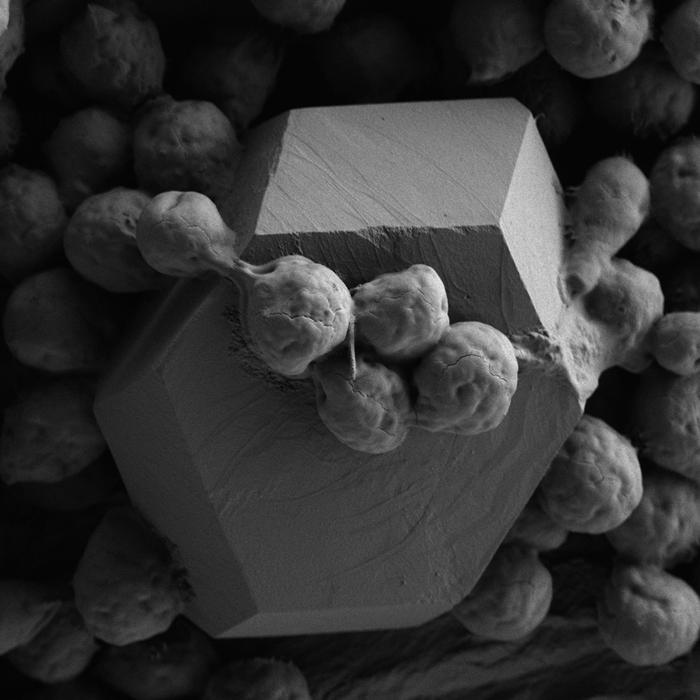
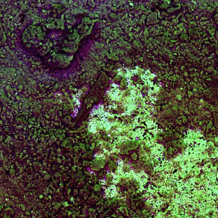
MultiDetector™ + Axial + Chamber BSE: each detector sits at a different take-off angle, each captures a different contrast.
Composite of graphene acid and zinc sulfide
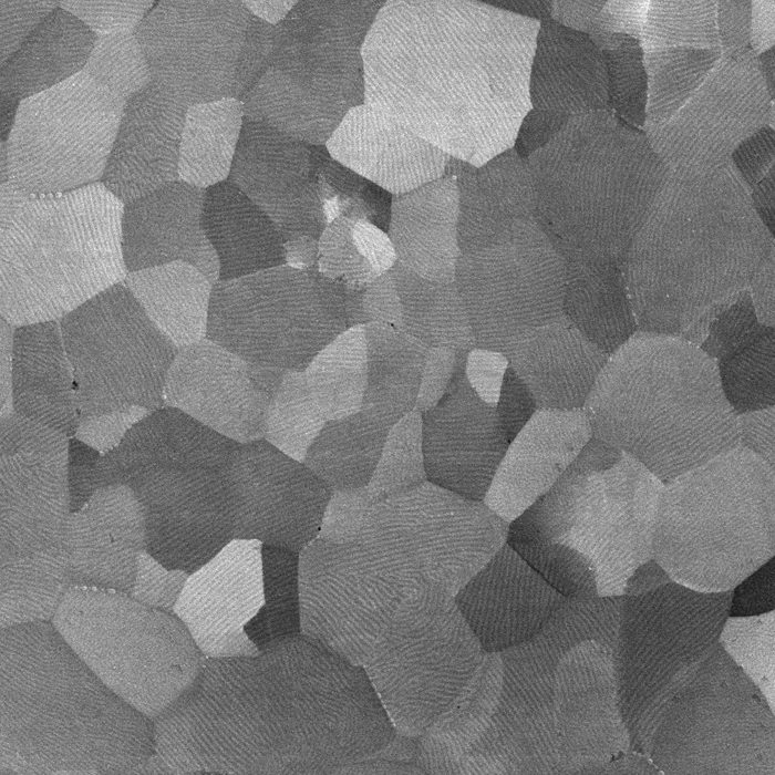
Field-free objective images magnetic samples without distortion; steels, NdFeB, and soft alloys at full resolution.
NdFeB magnet

1. Strong low-kV performance + field-free imaging

CLARA combines BrightBeam field-free optics with low-kV performance down to 500 eV, making it particularly well suited to surface-sensitive imaging and magnetic or beam-sensitive materials.

2. Multi-contrast BSE filtering

The combination of energy-filtered and angularly selective BSE detection is one of the strength of the new CLARA.
Instead of treating BSE simply as a compositional signal, CLARA provides greater control over which part of the BSE signal is detected, allowing researchers to emphasise different surface and material characteristics.

3. Reduced need for repeated imaging

Simultaneous SE and BSE acquisition can allow researchers to compare surface and material information from the same ROI without repeatedly repositioning the sample. This could be particularly valuable for quantitative and correlative workflows.

4. Very broad analytical integration

CLARA has been designed with 20 chamber ports and an open analytical architecture supporting techniques ranging from EDS and EBSD to Raman, WDS, ToF-SIMS and 4D-STEM.
That makes it an attractive platform for research groups that expect their analytical requirements to evolve.

5. Challenging-sample capability

The combination of low landing energy, field-free optics and MultiVac operation gives CLARA a strong proposition for non-conductive, magnetic, porous and beam-sensitive materials.

6. Workflow and automation

TESCAN has put significant emphasis on getting users from sample → ROI → optimised imaging → analysis quickly.
Wide Field Optics, automated alignment, In-Flight Beam Tracing, collision modelling and VisualCoder all contribute to this workflow.

  • All
  • AFM/SPM/SNOM
  • CL
  • CLEM
  • Diffraction Imaging
  • EBSD
  • EDS
  • Electron Beam Lithography (EBL)
  • Electron Microscopy
  • Fabrication
  • FIB
  • Hyperspectral
  • In situ
  • Micro XRF
  • Microscopy
  • Raman
  • SEM
  • Spectroscopy
  • TEM
  • Thermal Probe Lithography
  • WDS