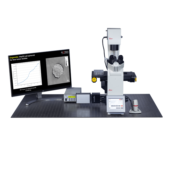
GLIM is an upgrade for your existing microscope that enables label-free, quantitative imaging for live cells, assays, tissues and organoids.
-
Key Features
- Quantitative Phase Imaging - Measure thickness and dry mass in large samples, non-invasive: no sample preparation.
- Designed for large specimens - Resolves samples with thickness from 50 µm – 350 µm+
- Multidimensional imaging at high speed - Up to 4D imaging (multichannel, time-lapse, Z-stack, tiling) at up to 12 fps
- Multichannel imaging - Seemless overlay of other microscope channels (ie fluorescence).
- Programmable 2D and 3D scanning - Using the microscopes full range of motion
- Integrates with existing microscopes - All major brands
-
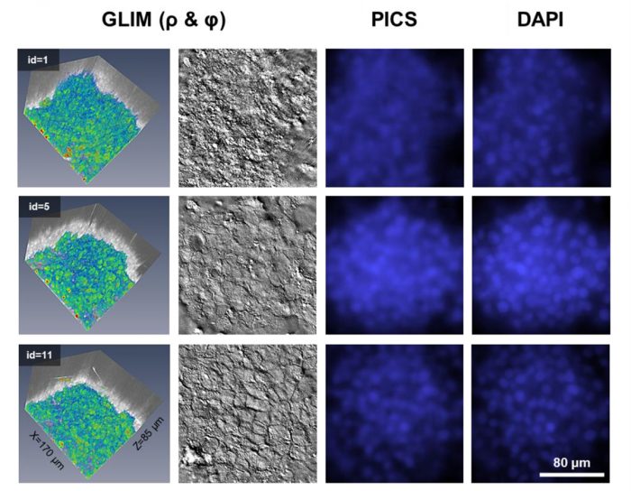
Phase Imaging with Computational Specificity (PICS)
Phase Imaging with Computational Specificity (PICS) is a new feature of Phi Optics QPI instruments CellVista GLIM™ and CellVista SLIM™ modules that enables various 2D (monolayer) and 3D (organoids) assays to have the accuracy and specificity of regular fluorescence but without the inconveniences associated fluorescent imaging of phototoxicity and photobleaching.
Click here to learn more about PICS.
Details about this new imaging capability was also published in Nature.
-
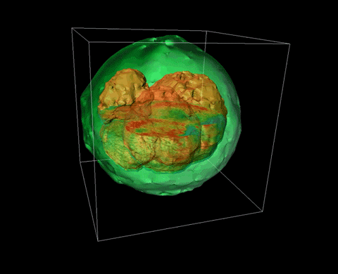
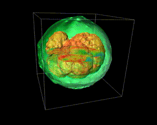
Bovine Embryo
Bovine embryo imaged using Phi Optics gradient light interference microscopy CellVista GLIM™.
- All
- Biological Microscopy
- Bioprinting
- Cell Culture
- Confocal Microscopy
- Digital Microscopy
- DNA/RNA
- Electron Microscopy
- Fluorescent Microscopy
- Light Microscopy
- Live Cell Imaging
- Microfluidic Bioprinters
- Microscopy
- Molecular Biology
- Multiphoton Microscopy
- Optical Tweezers
- Pre-clinical Imaging
- Protein
- Quantitative Phase Imaging
- Small Molecule
- Super Resolution Microscopy
- TEM
- Tomographic Microscopy