New AI-powered analysis function for SLIM and GLIM
Phase imaging with computational specificity (PICS) is a new feature (arriving in 2021) of Phi Optics QPI instruments that enables various 2D (monolayer) and 3D (organoids) assays to have the accuracy and specificity of regular fluorescence but without the inconveniences associated fluorescent imaging of phototoxicity and photobleaching.
More details about this breakthrough imaging modaility have been published in Nature. Read the paper here.
How Does PICS Work?
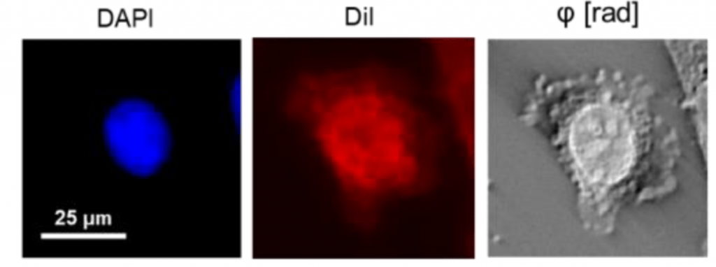
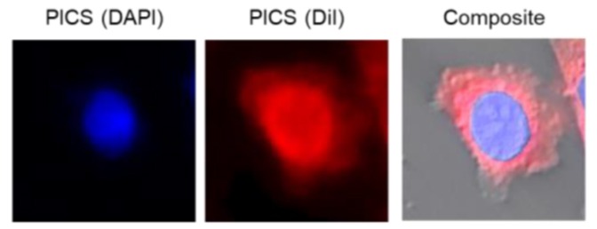
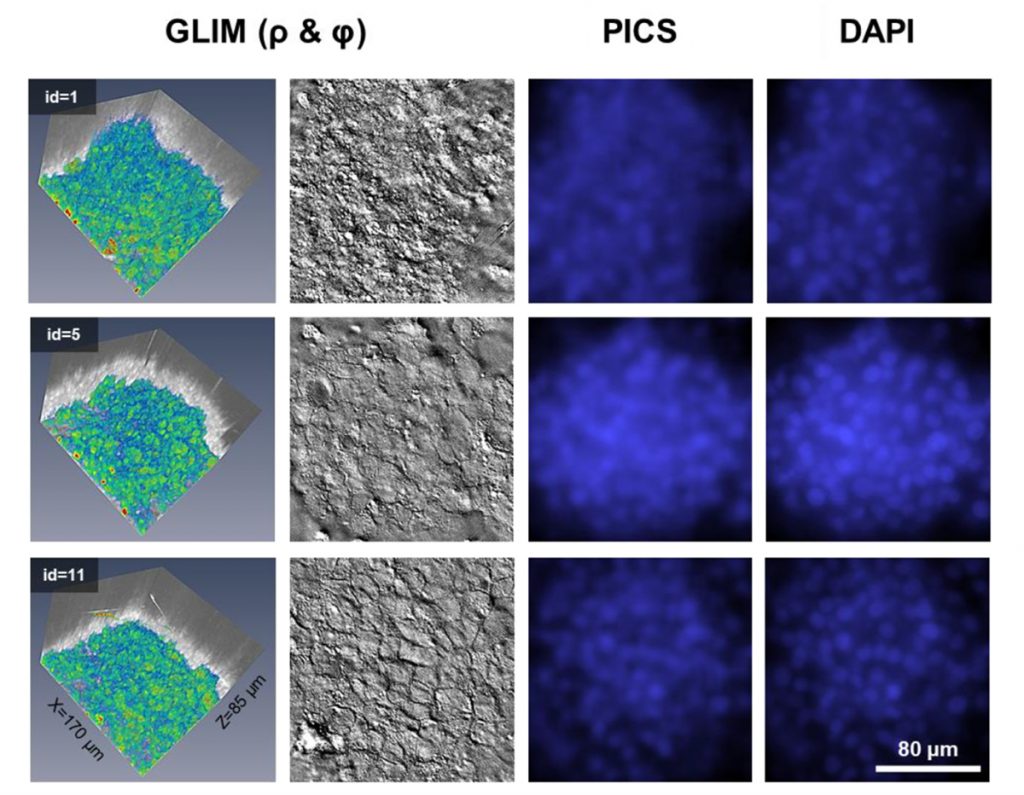
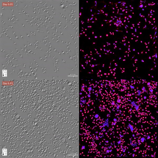
In PICS, a neural network learns from label-free (Phi Optics SLIM or GLIM) and reference fluorescence images to predict where specific fluorophores would bind in an unlabelled specimen. This enables (label-free) digital staining of cells using Artificial Intelligence (AI) and Deep Neuronal Network (DNN). The accuracy of the PICS prediction is made possible by SLIM and GLIM nanometre sensitivity combined with high signal to noise ratio as compared to classical phase (PC, DIC) or other quantitative phase techniques.
Assays
The specificity masks obtained from the computations are used back in the SLIM or GLIM maps to extract morphology, protein content and refractive index information that are used in the following assays:
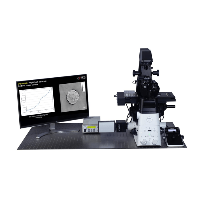
- Cell and spheroid counting, size, and 3D morphology, dry mass per cell and population, including time dependent statistics per cell and population
- Proliferation – label-free measurement of cell counts and dry mass (per cell and per population) as a function of time and growth inhibitor dose
- Cytotoxicity – label-free measurement of cell counts and dry mass (per cell and per population) as a function of drug dose
- Viability and Immune Cell Killing
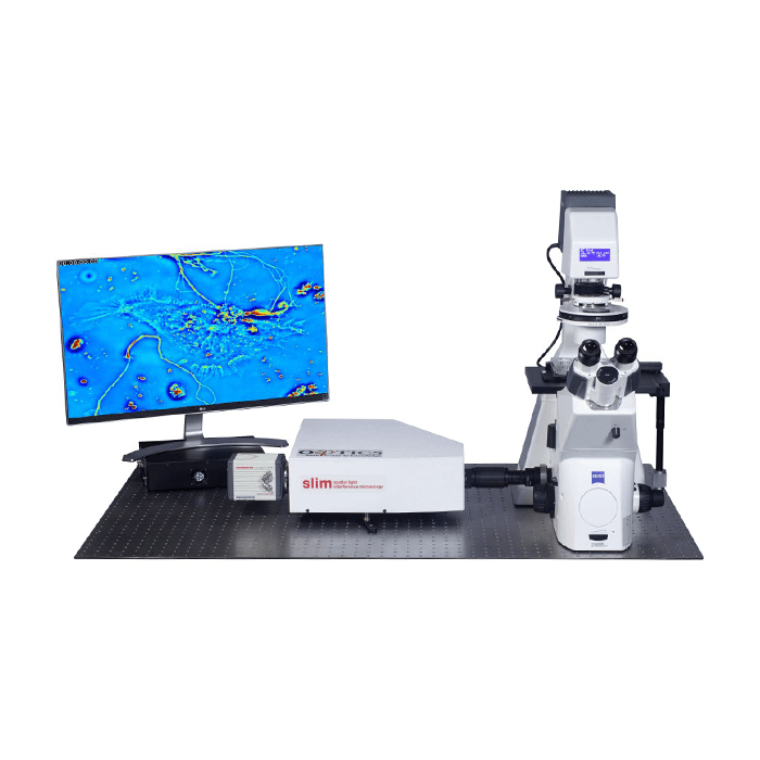
Upgrades for Existing Light Microscopes
Phi Optics SLIM and GLIM modules are available as upgrades for existing microscopes. The give your inverted light microscope analytical capabilities and attach to the camera port. Phi Optics CellVista software platform controls the motorisation of the microscope to deliver fully automated high-throughput multichannel acquisition. This combination results in seamless SLIM/GLIM + FL overlays for automated AI learning, without the need for manual annotation. Inference algorithms are loaded in CellVista software for live, continuous digital staining visualisation.
Human Epithelial Colon Cells
Liver Cells
Time Lapse Study
Related Products
GLIM
Gradient Light Interference Microscopy
SLIM
Spatial Light Interference Microscopy