CellVista SLIM™

Acquire mass and volume information in real time with label-free, quantitative imaging for live cells, assays, tissues and organoids.

Quantatative Phase addon for your inverted microscope, extract high resolution quantitative data from unstained and untreated cells over 2D, 3D and 4D.

  • Key Features

    • Real-time quantitative phase imaging and display. Watch the data acquisition scan as its happening
    • Label-free, low-illumination power capable. Non-destructive, excellent for long-term live cell, tissue and assay investigation
    • Programmed 2D and 3D scanning in a large field of view. See large surface areas and capture mass, volume and surface area, tracking the evolution of cells and tissues over time
    • Seamless overlay with other microscopy channels. All images captured on the same camera – great compliment for fluorescence and other channels
    • Auto segmentation tool. Allows for quick and easy sorting

The CellVista SLIM™ module is connected to the imaging port of a Phase Contrast microscope, with a camera connected at its output. The microscope separates the illumination into a sample and a reference beam that pass through the sample and are collected at the imaging port with a phase shift between them. The beams pass through the CellVista SLIM™ module where an electro-optical system introduces four accurately controlled phase delays between them. The CellVista SLIM™ camera acquires an intensity image for each phase delay. The intensity images are combined by interference and a recombination algorithm outputs the quantitative OPL map of the entire field of view of the microscope objective. The OPL map is converted to specimen height/volume, dry mass, and refractive index.

  • What is Quantitative Phase Imaging (QPI)?

    Optical path length (OPL) changes are subtle differences in thickness and refractive index in a transparent object. Quantitative Phase Imaging (QPI) refers to a group of techniques that include Spatial Light Interference Microscopy (SLIM) and Gradient Light Interference Microscopy (GLIM). SLIM and GLIM measure the OPL changes introduced by the sample and record it as pixel values in the generated image. Pixel intensity is proportional to the optical path length (OPL) change and enables direct measurement of the morphology, refractive index and dry mass of the object.

    SLIM and GLIM combine phase imaging with low-coherence interferometry and holography in a common-path geometry. This provides high signal to noise ratio (nanometer phase sensitivity) and low noise floor (temporal stability) when compared to Phase Contrast (PC), Differential Interference Contrast (DIC) or other QPI methods. SLIM and GLIM use the white light illumination of the microscope which avoids the speckles that plagues laser illumination-based QPI techniques and improves the optical sectioning due to the low-coherence length of the light source. Measurement throughput is high (up to 100 microliters measurement volume per minute) while simultaneously providing submicron resolution at high sample density (up to 10^10 particles/mL).

    SLIM and GLIM were invented at University of Illinois at Urbana Champaign by the Professor Gabriel Popescu QLI group. Phi Optics is commercializing SLIM and GLIM as add-on modules for commercial PC and respectively DIC microscope frames in a 4f-optical relay design that provides high-fidelity optical field reconstruction between the object and image planes with near diffraction-limited operation.                   

  • Phi Optics Phase Imaging with Computational Specificity (PICS)

    Phase Imaging with Computational Specificity (PICS)

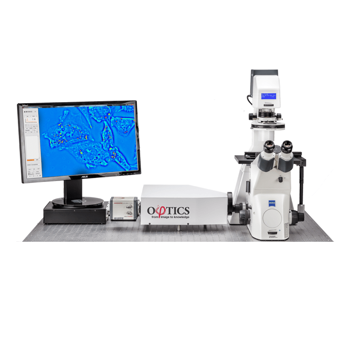
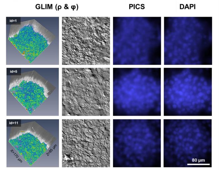
    Phase Imaging with Computational Specificity (PICS) is a new feature of Phi Optics QPI instruments CellVista GLIM™ and CellVista SLIM™ modules that enables various 2D (monolayer) and 3D (organoids) assays to have the accuracy and specificity of regular fluorescence but without the inconveniences associated fluorescent imaging of phototoxicity and photobleaching.

    Click here to learn more about PICS.

    Details about this new imaging capability was also published in Nature.

  • Fast detection of COVID-19 using light microscopy - SLIM from Phi Optics - imaging data

    Detecting COVID-19 Using CellVista SLIM™

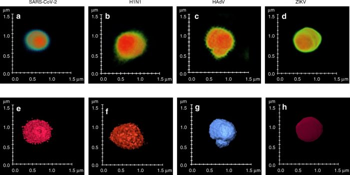
    Researchers from the Beckman Institute, the University of Illinois Urbana-Champaign, and the University of Illinois at Chicago lead by Professor Gabriel Popescu have devised an innovative label-free approach, using CellVista SLIM™ and Artificial Intelligence (AI) that was shown to be 96% successful in distinguishing the SARS-CoV-2 virus from other virus’ in preclinical testing. Their research was published in the prestigious journal Nature.

    Read more

  • All
  • Biological Microscopy
  • Bioprinting
  • Cell Culture
  • Confocal Microscopy
  • Digital Microscopy
  • DNA/RNA
  • Electron Microscopy
  • Fluorescent Microscopy
  • Light Microscopy
  • Live Cell Imaging
  • Microscopy
  • Molecular Biology
  • Multiphoton Microscopy
  • Optical Tweezers
  • Pre-clinical Imaging
  • Protein
  • Quantitative Phase Imaging
  • Small Molecule
  • Super Resolution Microscopy
  • TEM
  • Tomographic Microscopy