
CellVista GLIM™
Biological MicroscopyLight MicroscopyLive Cell ImagingQuantitative Phase Imaging
Quantitative Phase Imaging for Embryos, organoids, spheroids and other large samples.

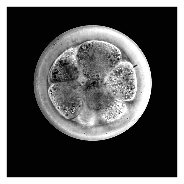
This Application Note illustrates the use of Gradient Light Interference Microscopy (GLIM) to image optically thick specimens such as tissues, organoids, embryos, and model multicellular organisms. GLIM rejects much of the multiple scattering contributions and yield high contrast these specimens. Furthermore, the illumination condenser aperture is fully open, which lends GLIM very strong optical sectioning so GLIM can provide 3D tomographic imaging.
Phi Optics CellVista GLIM units are modular and can upgrade any commercial inverted DIC microscopes thus allowing easy overlay and pixel registration with fluorescence channels. GLIM can measure the dry mass, refractive index and morphology over spatial scales from micrometers to millimeters, and temporal scales ranging from milliseconds to weeks. GLIM has applications in areas such as oncology, ART/IVF, crop sciences, developmental biology and in drug discovery/safety.