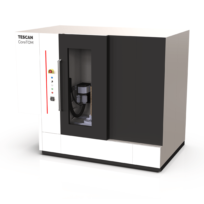
CoreTOM is a multi-resolution 3D X-ray microscope optimized for high resolution, large field of view imaging of full cores down to microplugs
Key Features
Multi-scale imaging Handles full size cores (Up to 1m tall) to microplugs with resolutions down to 3 μm.
Fast scanning & high sample throughput High power X-ray source with various signal-to-noise optimi¬sation decreasing scan time without sacrificing image quality
Intuitive volume of interest selection Real-time zooming in on selected regions based on the overview image
Dynamic in situ imaging Integration of in situ stages, provisions for flow and sensor lines and dedicated 4D acquisition and reconstruction protocols enable fast dy¬namic imaging
In situ integration kit Comprehensive hardware and software tools to facilitate the installation and control of peripheral equipment
Advanced sample mounting kit Large base plate with a grid pattern for mounting various setups, Clamp system for large samples, Kinematic mount plates. allowing max reproducibility and automated image registration, Gutter pattern to contain the spillage of fluids during in-situ experiments
MicroCT image of an elongated microplug of the Savonnières Limestone (FR). Using the automated stacked scan acquisition and merging tools in the CoreTOM microCT system, more representative data of elongated core samples can be obtained without compromising resolution.
X-ray CT image of a fossil mammoth tooth.
High resolution images inside the fossil can be obtained without physical sub sampling using the VoiS feature
Dynamic in-situ imaging of solute transport in a porous limestone.
Pore scale visualization of solute transport of tracer salt CsCl (10 wt.%) injected at a constant flow rate of 0.25 μl/s.
3D mapping of preferential flow paths (advection controlled) and more stagnant pore bodies (diffusion controlled).


")


