
X-ray diffraction (XRD) is a staple tool for materials identification in materials science. It is commonly used to determine the phase assemblage of crystalline materials, i.e. for materials like Silica (SiO2), a number of polymorphs exist including quartz, cristobalite and tridymite, with XRD being able to distinguish their presence based on the differing crystal structures, despite their common chemical composition.
How Does X-Ray Diffraction Work
A crystal structure is defined as a highly ordered, regularly repeating structure at the atomic or molecular level, forming symmetric patterns. These structures are 3-dimensional, with the smallest repeating segment being known as a unit cell. The size of the unit cell is dependent on the atoms involved (chemical composition) and the shape of the unit cell and is thus, unique to each phase. A more details explanation of crystal structures can be found elsewhere.
The theory of x-ray diffraction was developed by Von Laue and Bragg in the early 20th century. It is based on the interference phenomena between crystal structures and x-rays. If we consider a parallel beam of x-ray impinging the sample, they will see crystal lattices arranged in planes. Based on the spacing between these planes, at certain angles the reflected x-rays beams will undergo “constructive interference”, which can be seen as peaks in the diffraction pattern. These instances will only coincide with when the path difference between the x-ray beams is an integral number of wavelengths apart. By using monochromatic x-rays (i.e. x-rays of a single wavelength such as those generated by a copper anode), the wavelength is known and hence lattice parameters can be calculated according to Braggs law:
n.λ = 2d.sinθ
where:
n= integer (12,3…)
λ = Wavelength (nm)
d= Lattice spacing
θ = Diffraction angle
Amorphous materials have a disordered structure, i.e. no repeating segments as is the case with crystalline materials. As such they do not produce distinct peaks in a diffraction pattern. This can be seen in the comparison below that shows the diffraction pattern of a crystalline silica sample and an amorphous glass sample, that has a predominantly silica composition.
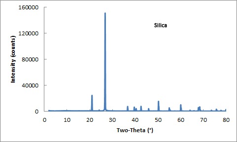
X-Ray diffraction pattern for silica (quartz), a crystalline material showing characteristic peaks.
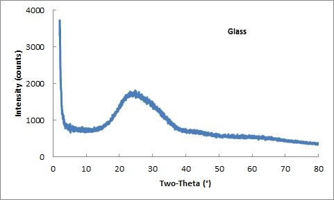
X-Ray diffraction pattern for glass, an amorphous material showing no distinct peaks.
Rigaku X-Ray Diffraction Systems
Rigaku are a leading producer of X-ray diffractometers with systems to suit all budgets and sample types.


Modern Diffractometers
Modern diffractometers vary in size and versatility. For routine XRD of such things as powders, benchtop systems are more than adequate, while larger floor standing systems offer the versatility of running different modes such as:
- Grazing Incidence XRD (GIXRD) – Suited to use with thin films, this geometry employs an x-ray beam fixed at a low angle, usually just above the angle for total reflection. This allows the maximum signal to be generated from the film, while minimal penetration is achieved, resulting in little if any signal being generated by the substrate.
- Transmission XRD – This involves passing the x-ray beam through the sample to produce a diffraction pattern. It is used for environmentally sensitive samples sealed in glass vials.
- Stress analysis (or Residual Stress Measurement XRD) – XRD can be used to measure the stress and strain (i.e. distortion) in a crystal lattice by comparing measured lattice parameters and comparing them to the known lattice parameters of the unstressed material.
- Small Angle X-Ray Scattering (SAXS) – This technique is used to measures the elastic scattering from inhomogeneities within a material over an angular range of ca. 0.05 – 5°. These scattering profiles can be used to determine information about the sizes and shapes of, and the distance and nature of the interactions between, these inhomogeneities.
- X-Ray Reflectivity (XRR) – This is a surface sensitive analytical technique used to characterise surfaces, thin films and multi-layers. It involves reflecting a beam of x-rays from a flat surface and measuring the intensity of the reflected x-rays in a particular direction (where reflected angle equals incident angle).
- Micro Area XRD or microdiffraction – This uses a highly focussed x-ray beam to analyse very small samples or small areas of large samples.
The key components of a diffractometer are described in the following sections.
X-Ray Generators
Rigaku also offer diffractometers that range in power from 300W to 18kW. Their lower power systems use conventional sealed x-ray tubes, while systems with power levels above 9kW, Rigaku’s patented rotating anode generator (RAG) technology is used.
Rotating anode technology is widely used with thousands of systems installed worldwide. RAGs are extremely reliable, with maintenance costs being comparable to lower powered sealed (X-ray) tube systems. The benefits of higher powered systems are, higher x-ray flux levels can produce data faster resulting in faster scans with the added ability of being able to resolve trace phases.
Read more about the Benefits of High Flux X-ray Diffraction for Materials Science.
Sample Stages and Loaders
Rigaku diffractometers have been designed to work with various sample stages and sample loaders to cater for various experiments.
To increase sample throughput, diffractometers can be fitted with sample carousels (with integrated spinner) or robotic magazine-based sample changes. These can be controlled through the software and allow users to load as many as 120 samples at once for measurement.
To cope with samples of various sizes and shapes, as well as different modes of measurement, a range of different stages are also available. These include:
- Sample spinners
- Environmental chambers for humidity, inert atmosphere, high temperature
- Battery cells
- Simultaneous XRD and DSC
XRD with DSC
Simultaneous collection of XRD and DSC data eliminates the uncertainty of separate measurements.
Mapping Stage
Allows xy positioning of the sample in the X-ray beam. 20 mm, 50 mm, and 75 mm translations available.
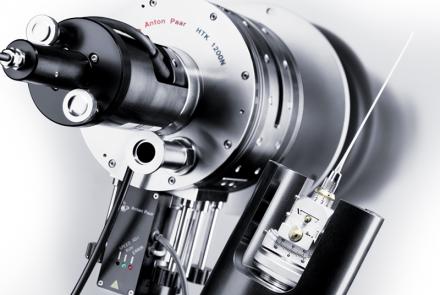
High Temperature Heating Chamber
High-temperature heating stage for powders and polycrystalline solid samples. Heating to 1200 °C in air and vacuum possible.
Detectors
Rigaku offer some of the most advanced and versatile detectors on the market.
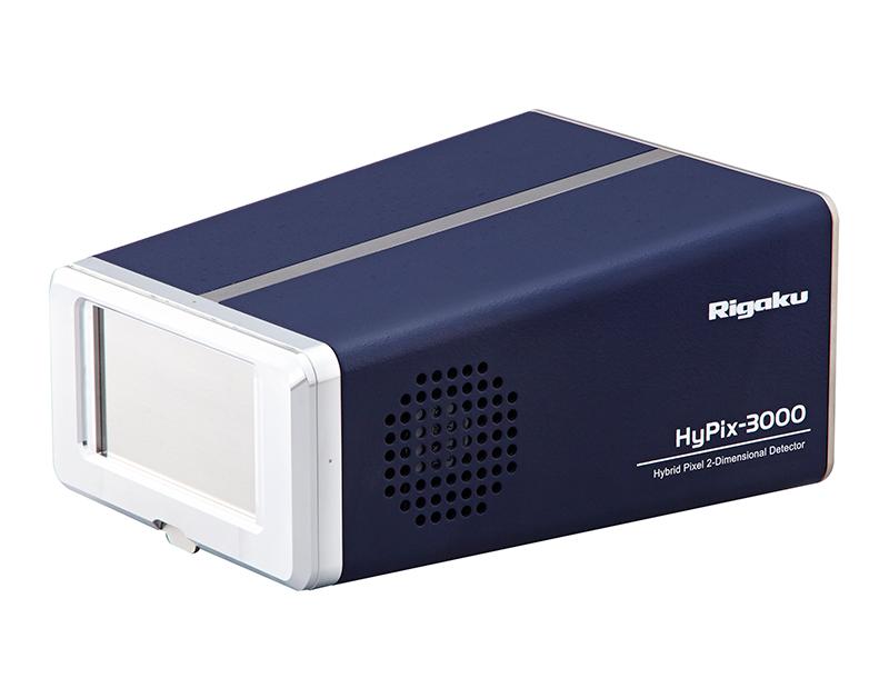
Starting with the HyPix-3000 Hybrid Photon Counting Detectors (HPC), which is a 2-dimensional semiconductor based photon counting detector with a large active area of approximately 3000mm2. It features a small pixel size of about 100µm square resulting in a detector with high spatial resolution. It also features a high count rate in excess of 106 cps/pixel, fast readout speed and effectively no background noise making it a high sensitivity detector with an ultra-high dynamic range. It can also be switched between 2-D, 1-D and 0-D modes seamlessly via the integrated software package with no hardware adjustments required achieving the design goal of optimal flexibility with minimal maintenance. Like its big brother the HyPix-400, it can be simply switched to the 0-D mode via the software and eliminates the requirement for an additional 0-D detector.
They also offer the D/tex Ultra 250, the world’s fastest 1-D detector. This model is based on semiconductor strip detector technology. Each detector is equipped with 256 strip channels, with each channel being 75µm wide and capable of count rates up to 106 cps and a global detector count rate of 2×10⁸ cps.
Read more about Hybrid Photon Counting Detectors.
XRF, The Ideal Companion for XRD
While XRD can be used to determine the phase assemblage of a material, it is very difficult to use it to determine the chemical composition of that material. E.g. if a mixture of sillimanite (Al2O3.SiO2) and Alumina (Al2O3) is analysed by XRD, it can certainly detect the presence of both phases. While XRD can be used to perform quantitative analysis to determine the relative amounts of each phase, it is unable to detect impurities, such as sodium or iron, which are detrimental if present in large enough proportions for refractory applications. XRF on the other hand can quite easily determine the relative amounts of Al and Si present, which can then be used to calculate the relative amounts of each phase (silliminate and alumina), while at the same time measuring the actual composition, including impurity levels.


