
With a long history in X-ray microscopy (XRM), Sigray, have recently launched the next generation PrismaXRM submicron 3D X-ray microscope that brings synchrotron-like performance to your laboratory. With industry-leading spatial resolution of 0.5µm for 3D tomography, voxel size below 60nm and the most advanced contrast imaging modes, you will be able to see things never before possible.
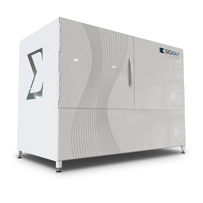
The PrismaXRM has already won the 2020 Microscopy Today Innovation award thanks to its flexible customisable platform. It incorporates the latest developments in x-ray technology, including a diamond backed transmission x-ray source, diffractive x-ray gratings and novel photon counting detector technology that take performance to the next level.
Sigray has revolutionised XRM with tri-contrast imaging. While X-ray absorption contrast microscopy has come a long way in recent times, there are still features and details that it can’t detect. Sigray now offer two addition imaging modalities that are simultaneously acquired.
- Quantitative Phase™ – a completely new phase contrast mode that provides quantitative access to refractive index and compositional information
- Subresolution Darkfield™ – reveals microstructural changes, including cracks and voids, that are otherwise invisible in absorption contrast
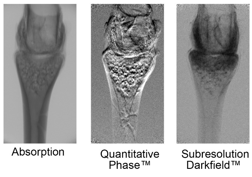
These new contrast imaging modes now allow you to see hidden defects (cracks and voids) and quantitative information on density to improve segmentation.
With these new imaging modes and resolution, the PrimsXRM is ideal for applications including:
- Materials – Carbon Fibre Reinforced Plastics (CFRP composites)
- Biomaterials – Unstained biological tissues (plants and animals)
- Energy – Batteries and fuel cells – in operando and in situ
- Failure Analysis – Cracks, voids, and delamination previously invisible for x-ray microscopy are now visible using the PrismaXRM’s unique Quantitative Phase and Subresolution Darkfield contrasts
- In situ Experiments – Three-phase flow, crack propagation, tensile and loading. Tri-contrast is particularly powerful for imaging in situ experiments.
If you have applications that you think would benefit from imaging using the new Sigray PrismaXRM, please contact us.