
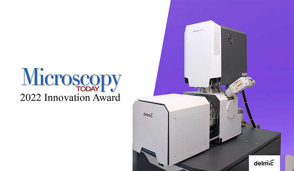
Congratulations to the DELMIC team following the announcement that FAST-EM was selected as one of the best microscopy innovations in 2022 by Microscopy Today, an esteemed journal in the field of microscopy.
Every year, Microscopy Today awards the ten most advanced technologies made in the field of light microscopy, electron microscopy (EM), and microanalysis. 2022 marks the fourth time of Delmic being the proud recipient of the Microscopy Today Innovation Award, following the successes of SECOM, DELPHI, and METEOR systems.
FAST-EM is an ultra-fast multi-beam scanning electron microscope (SEM) specifically designed and optimised for automating and speeding up the workflow of large volume EM for life science. The system was developed by a consortium, consisting of Thermo Fisher Scientific, Technolution, Delft University of Technology (TU Delft), and Delmic.
Reliable and extremely fast, the system can acquire images up to 100 times faster compared with standard SEM and can image samples for three days unattended. FAST-EM integrates a storage and data platform that allows an amount of data between 250 TB and 2000 TB to be locally stored. Furthermore, the platform provides an open interface to software that can be used to easily connect FAST-EM output to data-analysis and data-visualisation engines. With all those features, we can proudly say that FAST-EM introduces a revolution in EM workflows. For the first time, the entire workflow, from sample collection to insights, is harmonised seamlessly in one single solution.
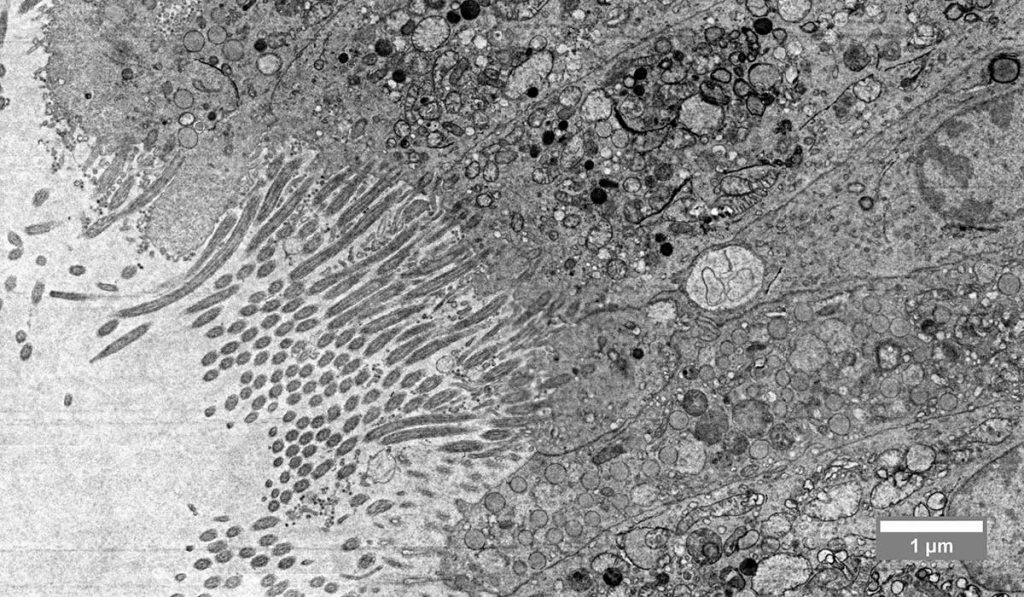
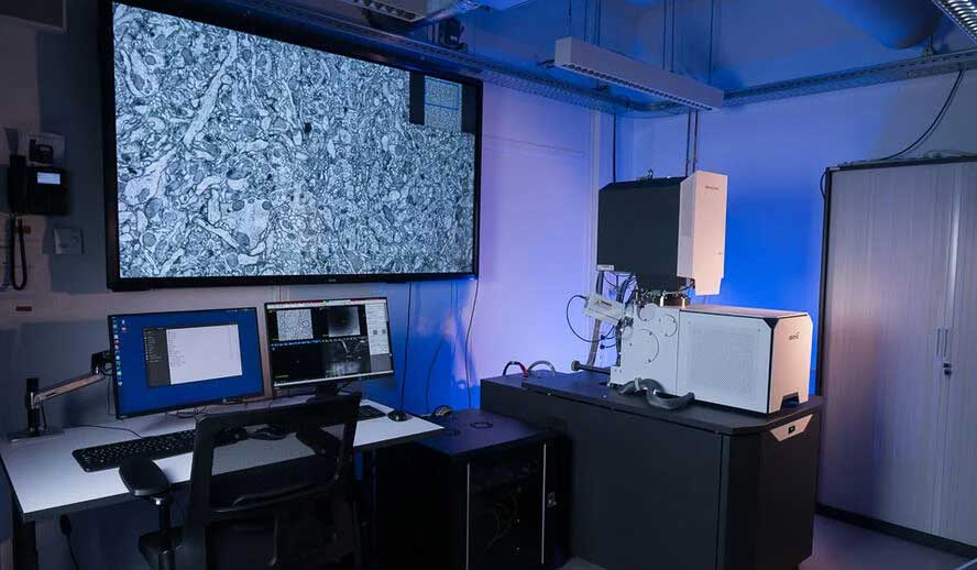
FAST-EM can reveal the ultrastructure of subcellular details over a large area of tissue, organ, or organism. It is extremely beneficial for large-volume 3D imaging, large-scale 2D imaging, and, in general, as a tool that can significantly speed up daily microscopy facility work. With FAST-EM, researchers and principal investigators now have a powerful tool that allows them to study complex and large biological samples, thus, answering their scientific questions in neurobiology, cell biology, histology, plant biology, pathology, biomaterials, and many more.
This award marks a great milestone for the Fast Imaging team and partners in their journey to unlock new areas for life science research that would otherwise be impractical!