
Evaluation of cell viability is crucial for research into such things as drug development and the design of other therapies for treating cancer and other diseases. Traditionally, cells are stained with chemical reagents or fluorescent markers that are able distinguish live cells from dead cells. Despite the broad acceptance of the technique several problems exist. A recent paper published in Nature Communications outlines a label-free technique, PICS (Phase Imaging with Computational Specificity) that has been shown to be 95% accurate for cell viability studies.
Problems with Fluorescent Labelling of Cells
While fluorescent labelling enables live cells to be differentiated from dead cells, there are a host of problems that make this technique undesirable for rapid, non-destructive and long-term investigations:
- Exogenous labelling generally requires some incubation time to achieve optimal staining intensity making it unsuitable for rapid assessments
- Exogenous labelling is toxic to the cells
- This toxicity precludes the technique from long-term studies
PICS – A Label-Free Alternative to Fluorescent Staining
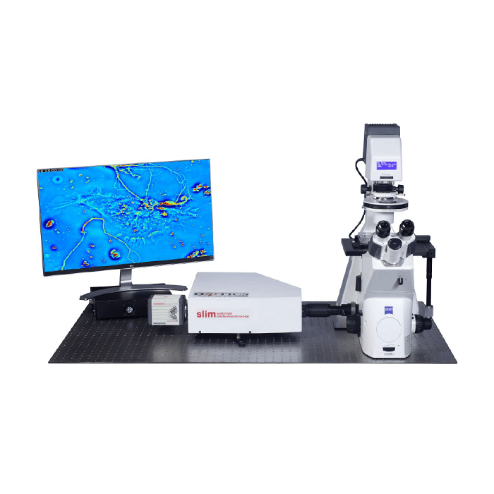 Quantitative Phase Imaging (QPI) is a label-free imaging modality that has been rapidly gaining acceptance in recent years. Spatial Light Interference Microscopy or SLIM is a highly sensitive QPI technique invented at University of Illinois Beckman Institute and commercialised by Phi Optics. SLIM measures optical path length changes (OPL) or phase shifts brought about by changes in thickness or refractive index of samples. It combines low-coherence interferometry with holography in a common-path geometry. Using broadband white light illumination it achieves sub-nanometer sensitivity to optical path length changes accompanied by high temporal stability. Using broadband illumination avoids speckles that plague laser-based QPI techniques as well as providing a low noise floor therefore a flat measurement background. Furthermore, the low power density of the white light overcomes phototoxicity facilitating long-term studies.
Phi Optics SLIM is supplied as a stand alone unit that can be easily retrofitted to phase contract microscopes using the C-mount making it compatible with most makes and models.
Using PICS to Assess Cell Viability
The technique described by the authors provides an instantaneous assessment of unlabelled cells using PICS to digitally stain live and dead cells. It involved using stained cells as a base line to collect fluorescence data which was then used to train a neural network which could then assess live, unstained cells.
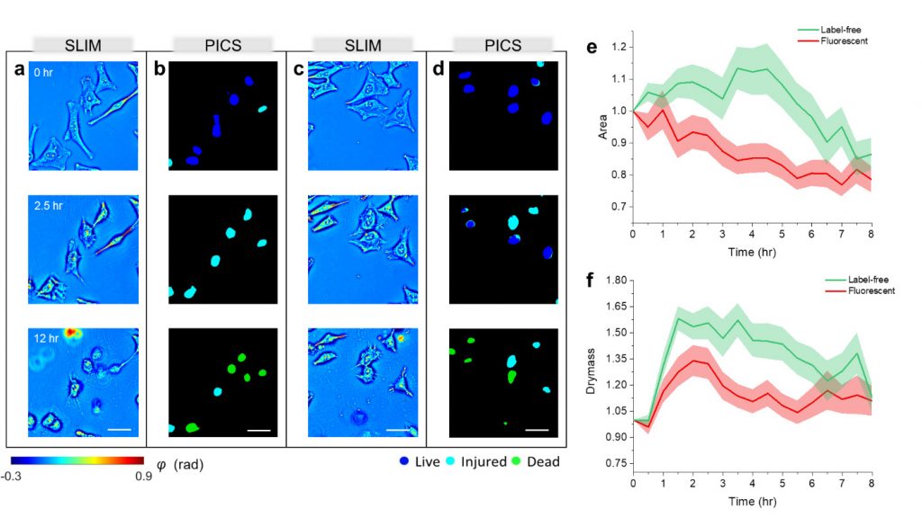
On tests performed on unlabelled HeLa cells and CHO (Chinese hamster ovary) cells, PICS was able to provide high-speed, label-free, unbiased viability assessment of adherent cells. It achieved this by using quantitative phase imaging to record high-resolution morphological structures of unstained cells which were assessed using deep learning techniques to extract intrinsic viability markets. This resulted in 96.7% and 94.9% accuracy respectively for HeLa and CHO cells.
Summary
PICS provides a rapid and accurate estimation of the viability of biological cells using a label-free technique, thus avoiding destructive fixation and staining procedures, while at the same time enabling long-term investigations. Cell viability is important for assessing the impact of drugs, physical or chemical stimulants, and other potential factors in cell function.
PICS can be easily added to existing inverted microscopes giving them additional QPI capabilities using the Phi Optics SLIM module.
Read the full paper “Live-dead assay on unlabeled cells using phase imaging with computation specificity” by Hu et al, in Nature Communications.