
3D Imaging of Optically Thick Specimens Such as Organoids, Tissue Assays, Embryos and Cell Clusters
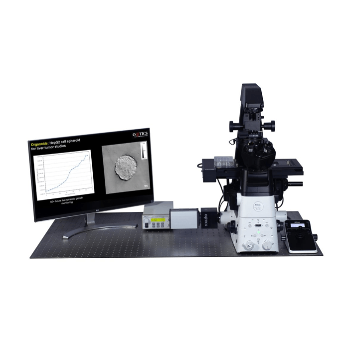
Phi Optics patented GLIM (Gradient Light Interference Microscopy) technology employs optical interferometry for extreme sensitivity to structure and dynamics. Phi Optics implements GLIM as an add-on to all major brand optical microscopes (10X to 100X magnifications). The GLIM approach to quantitative phase imaging provides speckle-free images due to high sensitivity of the measurement (nanometer scale spatial noise). Submicron optical sectioning is facilitated by high NA objectives and the micron-scale coherence length of the illumination – GLIM can render 3D tomographic images of transparent structures just by scanning the specimen through focus (Z-scanning).
What is GLIM?
Phi Optics GLIM is a non-invasive phase imaging technology that quantifies the optical path length differences in a biospecimen and converts them into thickness, dry mass area density and refractive index maps.
How Does GLIM Work?
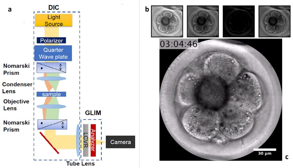
The principle of technology in its reference implementation is illustrated above. A biospecimen is imaged with the DIC modality of the microscope: two identical power, cross polarised beams are shifted transversely by the condenser (first) Nomarski prism with a shear distance smaller than the diffraction spot. After passing through the sample on the stage and the objective (second) Nomarski prism, the resulting interfering beams are sent into the GLIM module.
The GLIM module consists of a relay system including a liquid crystal variable retarder (LCVR) and a DIC analyser. The LCVR active axis is aligned to the polarisation direction of one beam and retards its phase by multiples of Pi/2 radians (quarter wavelength OPL) while the other beam is unmodified. By accurately controlling the phase shift between the two beams, we acquire multiple intensity (DIC) images at the camera plane, which have the same incoherent background, but different coherent contributions. Subtracting two pairs of images from each other (the sine and cosine components), we select only the coherent component and remove the multiple scattering contribution. The field interference equations are solved in each point of the frame and the result is a quantitative-phase image (QPI) that is uniquely determined and then integrated.
GLIM Features
- Live and time-lapse imaging
- Programmable 2D and 3D scanning using the microscope full range of motion
- Seamless multi-channel registration
GLIM and Fluorescence Microscopy
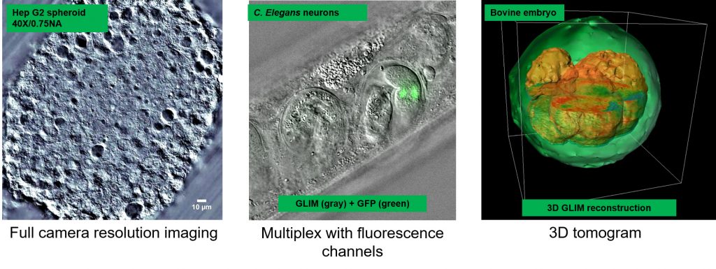
GLIM is a wide field quantitative imaging method. Thus, it can measure simultaneously multiscale fields of view at full camera resolution (e.g. 2 mm FOV for 10X objective at 4.2 MP camera resolution). Wide field optical sectioning (e.g. 850 nm Z-resolution for 100X/1.4NA objective) enables 3D tomography. All microscope output is acquired with the same camera which enables seamless overlay of GLIM images with fluorescence channels.
GLIM Applications
GLKIM can be added to most inverted research microscopes adding 4D label -free QPI (Quantitative Phase Imaging) capabilities. Examples of where it can be applied include:
- 3D morphology of embryos and cell clusters
- 3D organoid assays with single cell resolution
- Bioprinted cellular structures
- Animal and plant histology
- Microscopic animal models
If you only need to image thin specimens at high-resolution, you should look into SLIM – Spatial Light Interference Microscopy.