
On-demand webinar
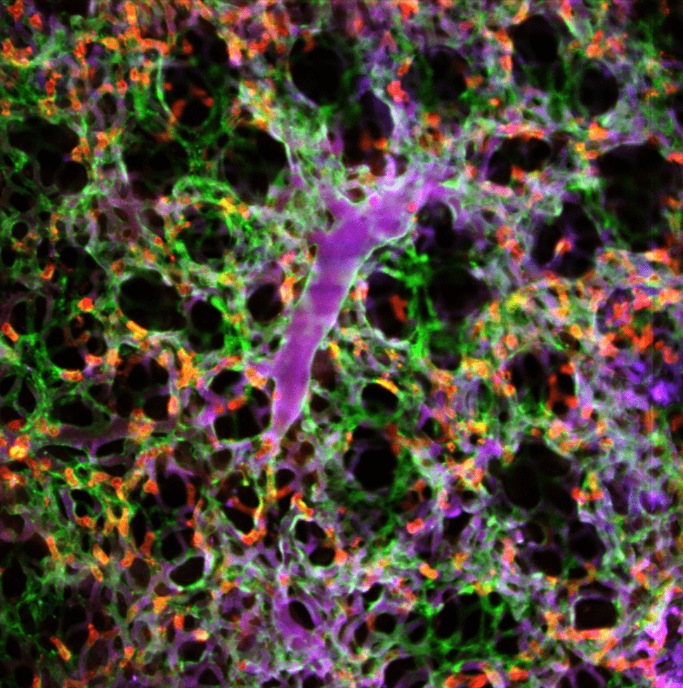
Dr Kim will present his work on a real-time laser-scanning intravital confocal/two-photon microscopy system extensively optimised for in vivo cellular-level imaging of internal organs in live animal models for various human diseases.
He will demonstrate how intravital microscopy can acquire real-time multi-color fluorescence microscopic images with sub-micron resolution. The special features of this system allow imaging of a wide variety of tissues and organs including:
- Skin
- Liver
- Pamcreas
- Kidney
- Heart
- Lung
- Small intestine
- Spleen
- Colon
- Retina
- Lymph node
- Bone marrow
Watch the webinar
We will email you details of how to access the webinar

Presenter – Dr. Pilhan Kim Ph.D.
Associate Professor, Korea Advanced Institute of Science and Technology, Korea
CEO, IVIM Technology, Inc., Korea
Dr. Pilhan Kim received B.S. and Ph. D. degree in Electrical Engineering from Seoul National University (Korea) in 2000 and 2005, respectively. From 2005 to 2010, he worked as a postdoctoral research fellow at Harvard Medical School, Massachusetts General Hospital Boston, USA, with cross-disciplinary postdoctoral fellowship from the Human Frontier Science Program (HFSP).
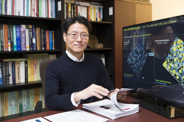
In 2010, he joined Korea Advanced Institute of Science and Technology (KAIST) as a tenure-track faculty member. Currently he is Associate Professor of Graduate School of Nanoscience and Technology at KAIST.
His main research interest is the development of an advanced in vivo cellular imaging technology based on ultrafast laser-scanning intravital microscopy systems. He developed this imaging system for systemic cellular-level visualisation of various preclinical model organisms to investigate the complex pathophysiology of human disease.