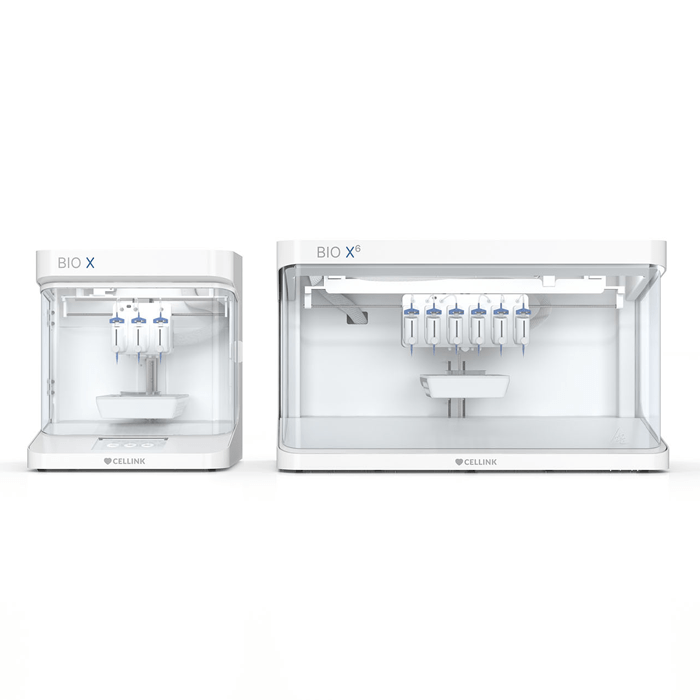
BIO X / BIO X6 - Extrusion Based Bioprinter
Cell CultureExtrustion Bioprinters

Three-dimensional cancer models such as spheroids, tumoroids and organoids have become common tools for preclinical drug development. However, a major limitation of these models is the lack of a native microenvironment where cell-cell communications and cellular migrations take place within an extracellular matrix (ECM)-based stroma. Establishing this native tumor microenvironment is critical for better understanding disease progression and developing robust drug discovery studies.
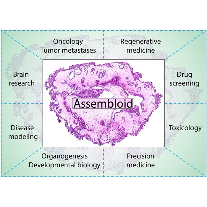
In the present study, a multicellular 3D bioprinted lung cancer assembloid model was developed by incorporating human lung cancer cells (A549), lung adenocarcinoma-associated fibroblasts and human umbilical vein endothelial cells (HUVECs) in a laminin-collagen-rich stromal environment. Using genetically encoded fluorescent cancer cells, the migration of cancer cells and merging of cancer spheroids within the matrix were visualised and further confirmed by histological analysis of fixed assembloids within the bioprinted constructs. Immunofluorescent staining was conducted to verify the expression of cell-surface and intracellular markers of 3D lung cancer tissue.
Furthermore, this study demonstrates that the embedded cancer cells expressed PD-L1, implying the suitability of such assembloid models for in vitro immunotherapy studies. The model and analysis protocols described here could be easily adapted for other cancer and healthy tissue models, in applications such as developmental biology, regenerative medicine and toxicology.
