
Nanoimager Super-Resolution Microscope
Biological MicroscopyConfocal MicroscopyFluorescent MicroscopyLive Cell ImagingSuper Resolution Microscopy
The complete package in super-resolution microscopy

Introduction
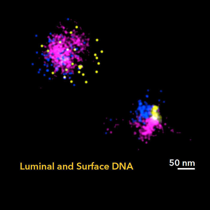
Extracellular Vesicles (EVs) are membranous particles that enable cell-cell communication via their surface components and cargo which includes proteins, lipids and genetic material such as DNA, mRNA and miRNA. While most of the transported material is carried within the lumen of EVs, a number of publications have identified a significant amount of genetic material coating the outer surface of vesicles (Figure 1). It is believed that the luminal contents of EVs are directly linked to their functional effect at recipient cells however, less is known about the role of DNA on the EV surface.
Studies have demonstrated that the presence of surface DNA modified EV adhesion properties1, contributed to the overall net negative charge of EVs2 and mediated horizontal DNA gene transfer3. Furthermore, on EVs derived from cancer cells, surface DNA contained oncogenic mutations that may imply a role of EV DNA in modulating the tumour microenvironment4. Therefore, spatial visualization of DNA and association with vesicles of discrete sizes or molecular signatures may contribute to a better understanding of the mechanisms and distinct functions of luminal and surface DNA in disease.