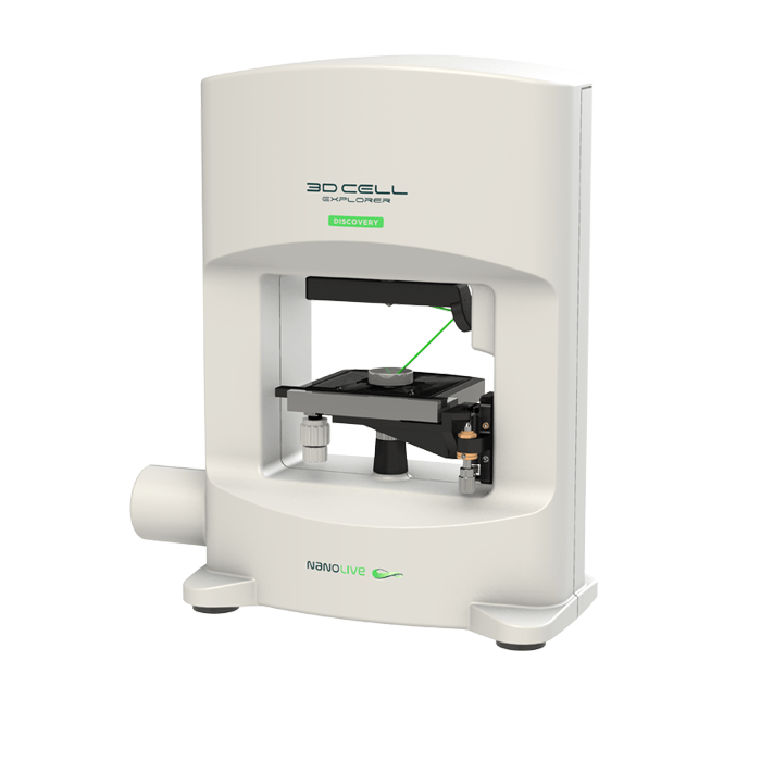
3D Cell Explorer - Live Cell Imaging Platform
Biological MicroscopyDigital MicroscopyLight MicroscopyLive Cell ImagingQuantitative Phase ImagingSuper Resolution MicroscopyTomographic Microscopy

In relation to the degree of dependency on the host cell resources, they can be classified in two different groups: facultative and obligate intracellular pathogens. Facultative intracellular pathogens like Listeria monocytogenes can survive both inside or outside cells, while obligate intracellular pathogens like Toxoplasma gondii need host cells in order to reproduce.
Host-pathogen interaction is the term used to describe how infectious agents survive within the host organisms.
In order to understand host-pathogen interactions at a cellular level, we need to overcome conventional microscopy methods limitations such as the impossibility to obtain 3D information or perform long-term live cell imaging avoiding phototoxicity and image quality loss.
Digital, marker-free, non-invasive imaging from Nanolive’s 3D Cell Explorer allows for a time-reduced sample preparation, high quality live-cell imaging at unique spatiotemporal resolution (<200nm; 1img/2sec) without any problem of photobleaching or phototoxicity.
Find further information and more examples on the following link:
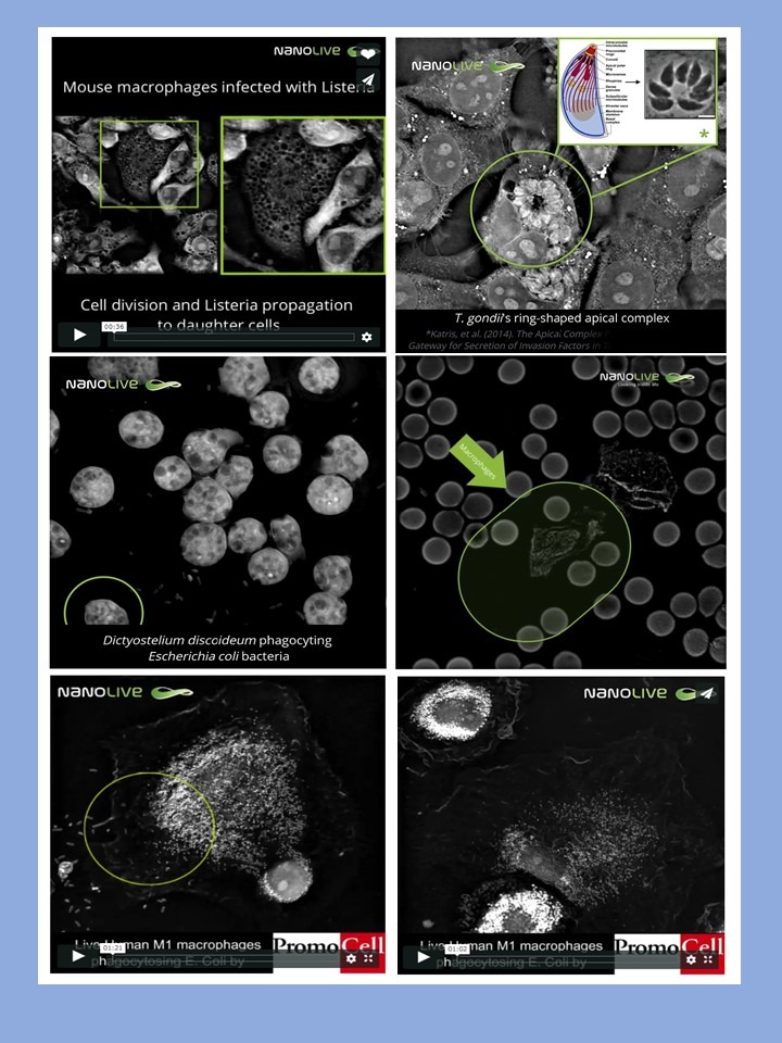
In this footage, we can distinguish T. gondii’s apical ring-shaped complex in tachyzoites from a Toxoplasma gondii infection in mouse fibroblasts (MEF1 T2). This structure plays a key role in host cell invasion processes. The 3D render obtained solely from the sample’s refractive index, an inherent characteristic from it (thus, from a non-stained sample) is compared to a static image obtained from confocal laser-scanning microscopy.
Under Nanolive’s 3D Cell Explorer, the identification of the parasitophorous vacuoles created during parasitic entry to the cell is possible, as well as a follow-up on the tachyzoites multiplication within the vacuoles. Thanks to the 3D Cell Explorer live cell imaging capabilities, the monitoring of host cell death and burst, followed by tachyzoites release can be observed.
