

3D Cell Explorer - Live Cell Imaging Platform
Biological MicroscopyDigital MicroscopyLight MicroscopyLive Cell ImagingQuantitative Phase ImagingSuper Resolution MicroscopyTomographic Microscopy
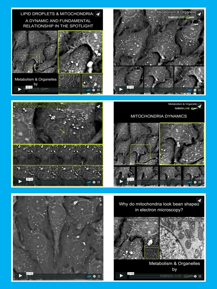
For the first time ever explore the inside of a living cell in 3D without the need for labeling. Take your Cell Research to the Next Dimension

