
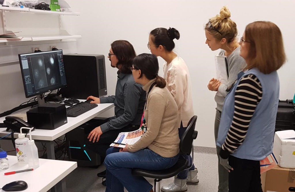
The recent installation of an Oxford Nanoimaging single molecule microscope at the Westmead Research Hub will provide researchers with much needed super resolution imaging capabilities. The microscope will allow researchers to observe cellular interactions at the nanoscale, giving them greater insights into how diseases such as cancer behave, increasing the chances of developing breakthrough cures.
The Nanoimager is a unique super resolution microscope that offers biological researchers a range of imaging modes including Direct Stochastic Optical Reconstruction Microscopy (dSTORM) Photoactivated Localisation Microscopy (PALM), Total Internal Reflection (TIRF), Highly Inclined and Laminated Optical Sheet (HILO) as well as Structural Illumination microscopy (SIM) in a single instrument. Other key design features include compact size, internal active vibration damping, a streamlined optical path and robust design to ensure the most stable of images are produced without the need for optical benches or dark rooms.
Dr. Laurence Cantrill, Advanced Microscopy and Imaging Specialist at Kids Research, the research arm of Sydney Children’s Hospitals Network said, “we have been interested in adding super resolution microscopy capabilities to our facility for some time. Solutions that we had previously looked at were usually too expensive. The Nanoimager was not only affordable, but also compact and future-proof. Its flexible design gives us the capability to add new imaging modalities down the track to cater for new research areas that we may not even be aware of yet, making it ideal for a multi-use facility such as ours. Evaluating the system last year, I was convinced that this microscope provided us with a solution that will suit the numerous and varied researchers in our organisation.”
The Nanoimager will be located at the Westmead Research Hub where it will serve researchers from nearby hospitals and research institutes, as well as the University of Sydney. “Having the instrument located locally is not only convenient, but also critical for many of the time-sensitive live cell studies that we carry out”, added Dr. Cantrill.
The user-friendly and robust design make it suitable for researchers from undergraduate to postdoctoral level and its simple but multimodal design makes it valuable for teaching the principles of sub-diffraction imaging. He also expects that it will be used by researchers in areas such as:
- Cancer research from its origins in telomere dysregulation through to its physiology and processes of invasion and migration
- Tracking the herpes simplex virus (HSV-1) in neurons
- Looking at damage and repair in cell membranes in muscular dystrophy
- Unravelling the early stages of HIV infection
Dr Cantrill is also grateful to the Ian Potter Foundation and a private philanthropist for their financial contributions that helped secure the Nanoimager.
AXT’s Managing Director Richard Trett also commented, “AXT have a proven track record of introducing new technologies to the Australian market. This is yet another example of how we are able to provide Australian researchers with the necessary tools to keep them at the forefront in their respective fields.”
The Oxford Nanoimaging single molecule microscope is part of AXT’s extensive microscopy and microanalysis portfolio that covers light microscopes, scanning electron microscopes (SEM), focused ion beams (FIB), detectors and associated sample preparation equipment. It includes systems that cater for industrial and high-end research applications across a vast array of scientific disciplines.