3D Cell Explorer-fluo – Live Cell Imaging

Holotomography Microscopy

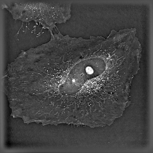
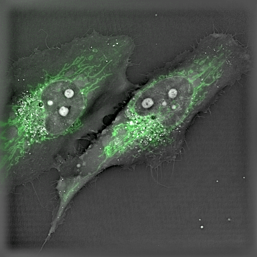
For the first time ever explore the inside of a living cell in 3D without the need for labeling. Take your Cell Research to the Next Dimension

  • Key Features

    • Resolution to below 200nm in X-Y - Image individual organelles
    • Full 3D scan in under 2 seconds - Fast imaging for capturing dynamic processes
    • Up to 4 correlative fluorescence channels - Covering the standard range of visible fluorophores for correlation of images
    • Long term live cell capable - Heat, humidity, CO2 and O2 control for long term stable timelapse imaging of even the most sensitive cells

The 3D Cell Explorer-fluo

A disruptive technology which, for the first time ever, allows users to explore the inside of a living cell in 3D without the need for any labeling or other invasive methods.

The 3D Cell Explorer-fluo is a high speed, high resolution and non-invasive tool that can look deep inside biological systems. This allows users to record stunning 3D images of entire cells in just seconds and with a higher resolution than any conventional light microscope on the market.

The latest version of the 3D Cell Explorer includes a three channel CoolLED PE-300 Ultra light source for rapid correlation of fluorescence probes in combination with the Tomo-Holographic image.

Multimodal complete solution for 3D live cell exploration: Combine 3D refractive index analysis with a fully integrated 3 channel fluorescence module to image your live cells as they are and as long as you want. Put chemical information into structural context for new biological insights.

Multiplexing: Transform 2D fluorescence into 3D cell tomography: Identify cell organelles through fluorescence and monitor non-invasively their structures and dynamics in 3D and marker-free. Explore fluorescence (3 channels) and Digital Stains (8 channels) simultaneously.

Extended live cell imaging: Image your live cells as long as you need. Limit cell damages caused by fluorescent markers, bleaching and phototoxicity.

  • A Self-Adjusting, Holotomographic Microscope

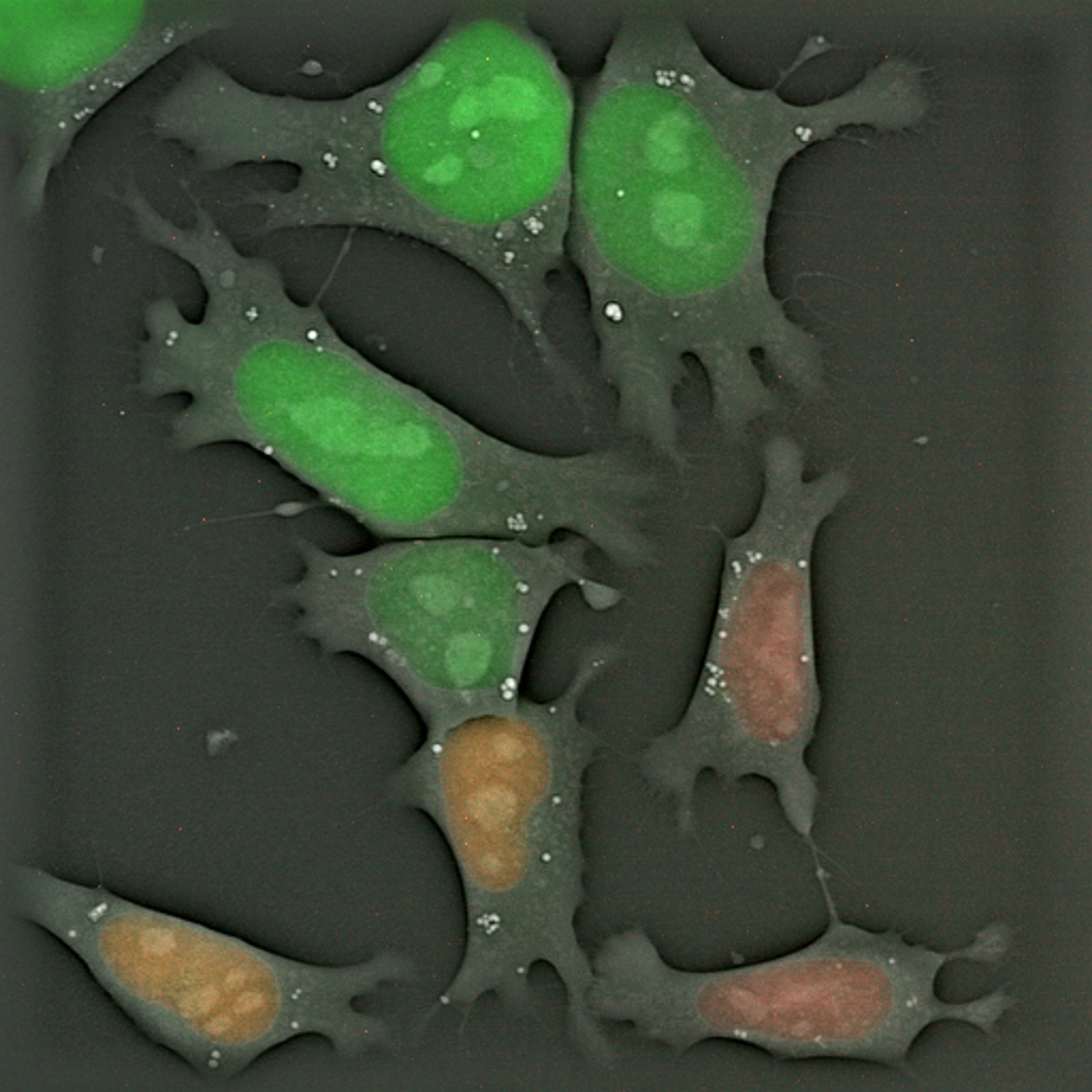
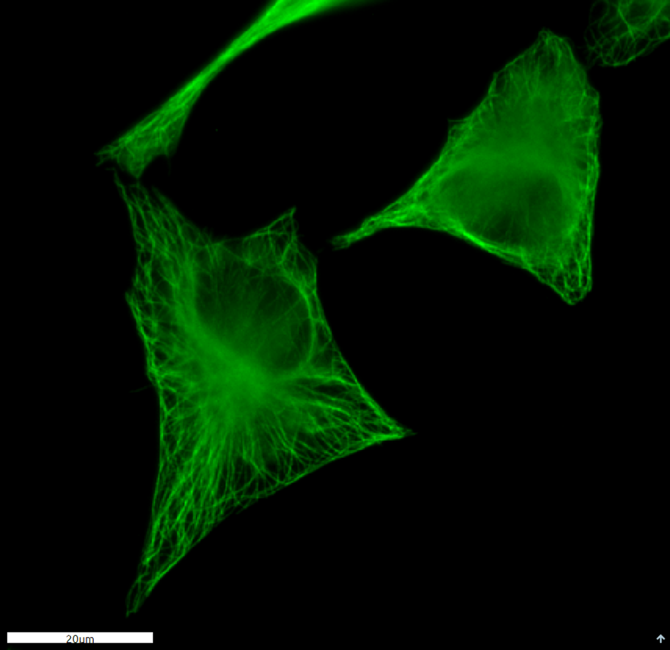
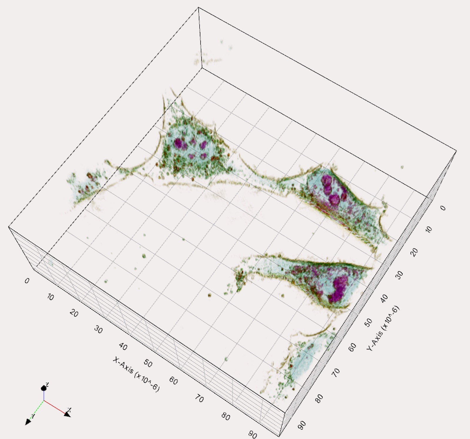
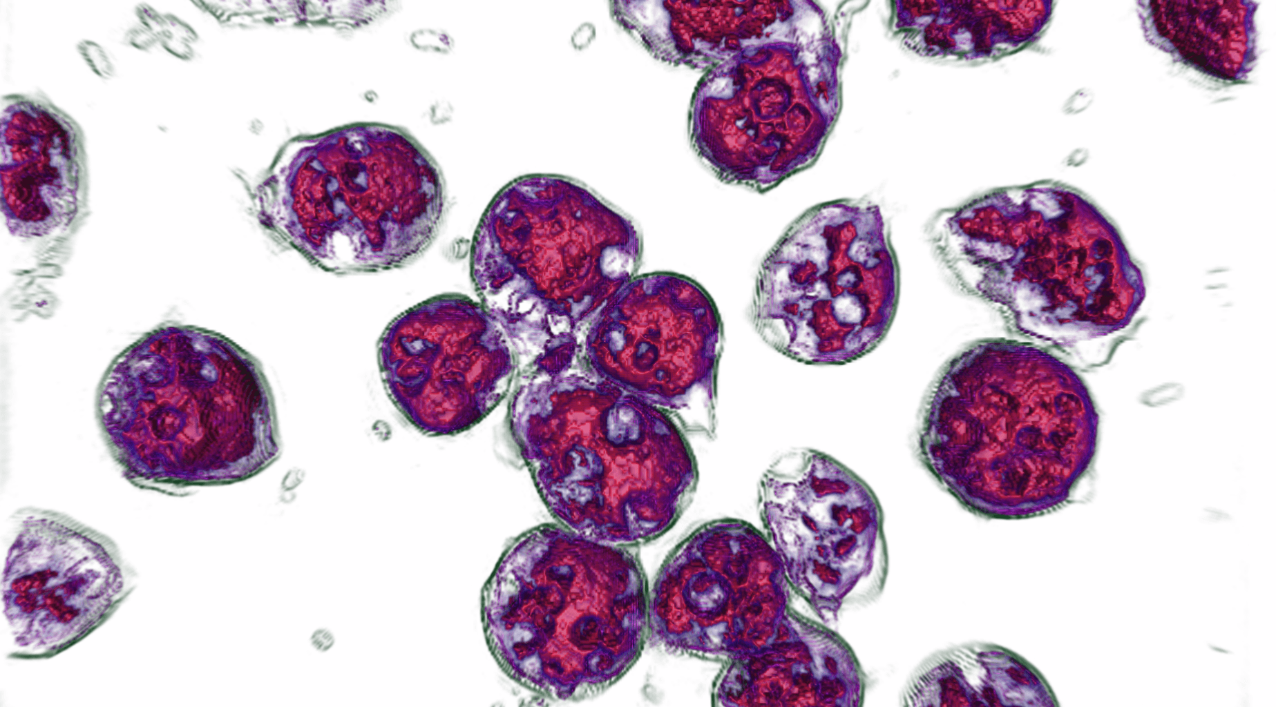
    This 3D holotomographic live cell microscope is the result of years of work and development. Through a combination of holography and rotational scanning the system detects changes to light as it propagates through the cell, i.e. the 4D distribution of the physical refractive index (RI) within the cell.

    The sample is positioned between a high-numerical-aperture air objective beneath the sample and a rotational illumination arm above. Green light (520 nm) from a laser diode is split into sample and reference beams. The sample is illuminated with a laser beam inclined at 45° which rotates around the sample 360°. A series of holograms is recorded on a digital camera by combining the beam that has passed through the sample with the reference beam.

  • STEM CELLS: Long Term Live Imaging of Mouse Embryonic Stem Cells for 48 hours!

    Live imaging of mouse Embryonic Stem Cells for 48 hours. 1 image every 15 seconds. This is unique material as never seen before.
    Technology: 3D Cell Explorer microscope from Nanolive, Switzerland.

    To see more Nanolive videos, please click here


  • Live FUCCI mESC were imaged for over 48 hours with the Nanolive’s 3D Cell Explorer

    A holotomographic image was taken every 14 seconds while a double channel (green and red) epifluorescence image was only taken every 30 holotomographic frames (7:30 minutes). The difference in acquisition rate was performed to reduce fluorescence related phototoxicity.

    *Please note that the latest fluorescence image is displayed over the following 30 holotomographic images until a new one is acquired, for visualization purpose. The mismatch between the two signals is due to this image processing choice.

  • Smart Lipid Droplet Assay

    Run time – 1:31 min

    The first smart digital assay to provide a push button solution for analysing lipid droplets dynamics, label-free.

  • All
  • Biological Microscopy
  • Bioprinting
  • Cell Culture
  • Confocal Microscopy
  • Digital Microscopy
  • DNA/RNA
  • Electron Microscopy
  • Fluorescent Microscopy
  • Light Microscopy
  • Live Cell Imaging
  • Microscopy
  • Molecular Biology
  • Multiphoton Microscopy
  • Optical Tweezers
  • Pre-clinical Imaging
  • Protein
  • Quantitative Phase Imaging
  • Small Molecule
  • Super Resolution Microscopy
  • TEM
  • Tomographic Microscopy