
Nanoimager Super-Resolution Microscope
Biological MicroscopyConfocal MicroscopyFluorescent MicroscopyLive Cell ImagingSuper Resolution Microscopy
The complete package in super-resolution microscopy

Extracellular vesicles (EVs) play key roles in cell-to-cell communication. EVs can cross biological barriers (such as the blood-brain barrier) and get internalized into the cell with a high degree of specificity.
Until recently, due to their size and design, the majority of super-resolution microscopes have not been able to support research in enclosed, ventilated biosafety cabinets. But things are changing.
Viral particles vary greatly in size, but are typically below the resolution limit of conventional light microscopy. Recently, super-resolution techniques have been employed to study their mechanistic and functional characteristics at a single-molecule level.
The Nanoimager can track single molecules and vesicles in both bacterial and eukaryotic cells with super-resolution microscopy.
Understanding and quantifying viral particle behavior.
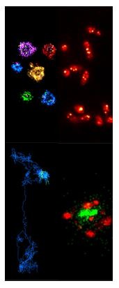
Gain detailed understanding of cellular features through multi-color super-resolution microscopy with advanced data analysis including colocalization and clustering.
Intensity measurements, super-resolution and multi-color labeling for characterizing protein complexes and their assembly.
A dynamic, real-time nanoscale ruler, now a general tool for characterizing molecular interactions and structure with Alternating Laser Excitation (ALEX) support.
DNA-PAINT provides easy localization-based super-resolution with nanorulers from GATTAquant GmbH.
Understanding the structure and function of extracellular vesicles through super-resolution microscopy.